 Movie
Movie Controller

[English] 日本語
 Yorodumi
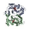
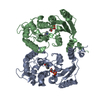
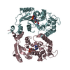
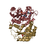
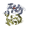
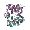
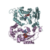
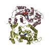
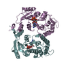
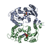
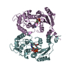
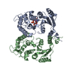
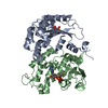
- PDB-3i9k: Crystal structure of ADP ribosyl cyclase complexed with substrate NAD -
+ Open data

- Basic information
| Entry | Database: PDB / ID: 3i9k | ||||||
|---|---|---|---|---|---|---|---|
| Title | Crystal structure of ADP ribosyl cyclase complexed with substrate NAD | ||||||
 Components Components | ADP-ribosyl cyclase | ||||||
 Keywords Keywords | HYDROLASE / Homodimer / protein substrate NAD complex / ADP-ribosyl cyclase / Disulfide bond / Fertilization / NAD | ||||||
| Function / homology |  Function and homology information Function and homology information2'-phospho-ADP-ribosyl cyclase/2'-phospho-cyclic-ADP-ribose transferase / phosphorus-oxygen lyase activity / NAD+ nucleosidase activity, cyclic ADP-ribose generating / Hydrolases; Glycosylases; Hydrolysing N-glycosyl compounds / single fertilization / transferase activity / cytoplasmic vesicle / plasma membrane Similarity search - Function | ||||||
| Biological species |  | ||||||
| Method |  X-RAY DIFFRACTION / SYNCHROTRON / MOLECULAR REPLACEMENT / Resolution: 1.83 Å X-RAY DIFFRACTION / SYNCHROTRON / MOLECULAR REPLACEMENT / Resolution: 1.83 Å | ||||||
 Authors Authors | Liu, Q. / Graeff, R. / Kriksunov, I.A. / Jiang, H. / Zhang, B. / Oppenheimer, N. / Lin, H. / Potter, B.V.L. / Lee, H.C. / Hao, Q. | ||||||
 Citation Citation | Journal: J.Biol.Chem. / Year: 2009 Title: Structural basis for enzymatic evolution from a dedicated ADP-ribosyl cyclase to a multifunctional NAD hydrolase Authors: Liu, Q. / Graeff, R. / Kriksunov, I.A. / Jiang, H. / Zhang, B. / Oppenheimer, N. / Lin, H. / Potter, B.V.L. / Lee, H.C. / Hao, Q. | ||||||
| History |
|
- Structure visualization
| Structure viewer | Molecule: MolmilJmol/JSmol |
|---|
- Downloads & links
-Download
| PDBx/mmCIF format | 3i9k.cif.gz | 122.7 KB | Display | PDBx/mmCIF format |
|---|---|---|---|---|
| PDB format | pdb3i9k.ent.gz | 94.6 KB | Display | PDB format |
| PDBx/mmJSON format | 3i9k.json.gz | Tree view | PDBx/mmJSON format | |
| Others |  Other downloads Other downloads |
-Validation report
| Arichive directory | https://data.pdbj.org/pub/pdb/validation_reports/i9/3i9kftp://data.pdbj.org/pub/pdb/validation_reports/i9/3i9k | HTTPS FTP |
|---|
-Related structure data
| Related structure data | 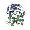 3i9jC  3i9lC  3i9mC  3i9nC 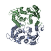 1lbeS C: citing same article ( S: Starting model for refinement |
|---|---|
| Similar structure data |


-Links
 PDBj
PDBj- Assembly
| Deposited unit | 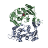
| ||||||||
|---|---|---|---|---|---|---|---|---|---|
| 1 |
| ||||||||
| Unit cell |
|
-Components
| #1: Protein | Mass: 29507.883 Da / Num. of mol.: 2 / Mutation: E179G Source method: isolated from a genetically manipulated source Source: (gene. exp.) Plasmid: PPICZ(ALPHA)A / Production host:  Pichia Pastoris (fungus) / Strain (production host): X-33 (INVITROGEN) / References: UniProt: P29241, NAD+ glycohydrolase Pichia Pastoris (fungus) / Strain (production host): X-33 (INVITROGEN) / References: UniProt: P29241, NAD+ glycohydrolase#2: Chemical | ChemComp-NAD / |   Mass: 663.425 Da / Num. of mol.: 1 / Source method: obtained synthetically / Formula: C21H27N7O14P2 / Comment: NAD*YM Mass: 663.425 Da / Num. of mol.: 1 / Source method: obtained synthetically / Formula: C21H27N7O14P2 / Comment: NAD*YM#3: Water | ChemComp-HOH / |  Mass: 18.015 Da / Num. of mol.: 461 / Source method: isolated from a natural source / Formula: H2O Mass: 18.015 Da / Num. of mol.: 461 / Source method: isolated from a natural source / Formula: H2OHas protein modification | Y | |
|---|
-Experimental details
-Experiment
| Experiment | Method: X-RAY DIFFRACTION / Number of used crystals: 1 |
|---|
- Sample preparation
| Crystal | Density Matthews: 2.41 Å3/Da / Density % sol: 48.94 % |
|---|---|
| Crystal grow | Temperature: 298 K / Method: vapor diffusion, hanging drop / pH: 7.5 Details: 0.1M imidazole, pH 7.5, 12-24% PEG 4000 , VAPOR DIFFUSION, HANGING DROP, temperature 298K |
-Data collection
| Diffraction | Mean temperature: 100 K |
|---|---|
| Diffraction source | Source: SYNCHROTRON / Site: CHESS  / Beamline: A1 / Wavelength: 0.9771 Å / Beamline: A1 / Wavelength: 0.9771 Å |
| Detector | Type: ADSC QUANTUM 210 / Detector: CCD / Date: Apr 1, 2007 |
| Radiation | Monochromator: Si 111 CHANNEL / Protocol: SINGLE WAVELENGTH / Monochromatic (M) / Laue (L): M / Scattering type: x-ray |
| Radiation wavelength | Wavelength: 0.9771 Å / Relative weight: 1 |
| Reflection | Resolution: 1.83→30 Å / Num. all: 49177 / Num. obs: 47800 / % possible obs: 97.2 % / Observed criterion σ(F): 0 / Observed criterion σ(I): 0 |
| Reflection shell | Resolution: 1.83→1.9 Å / % possible all: 84.1 |
- Processing
| Software |
| ||||||||||||||||||||||||||||||||||||||||||||||||||||||||||||||||||||||||||||||||||||||||||||||||||||||||||||||||||||||||||||||||||||||||||||||||||||||||||||||||||||||||||
|---|---|---|---|---|---|---|---|---|---|---|---|---|---|---|---|---|---|---|---|---|---|---|---|---|---|---|---|---|---|---|---|---|---|---|---|---|---|---|---|---|---|---|---|---|---|---|---|---|---|---|---|---|---|---|---|---|---|---|---|---|---|---|---|---|---|---|---|---|---|---|---|---|---|---|---|---|---|---|---|---|---|---|---|---|---|---|---|---|---|---|---|---|---|---|---|---|---|---|---|---|---|---|---|---|---|---|---|---|---|---|---|---|---|---|---|---|---|---|---|---|---|---|---|---|---|---|---|---|---|---|---|---|---|---|---|---|---|---|---|---|---|---|---|---|---|---|---|---|---|---|---|---|---|---|---|---|---|---|---|---|---|---|---|---|---|---|---|---|---|---|---|
| Refinement | Method to determine structure: MOLECULAR REPLACEMENT Starting model: PDB ENTRY 1LBE Resolution: 1.83→20 Å / Cor.coef. Fo:Fc: 0.958 / Cor.coef. Fo:Fc free: 0.93 / Occupancy max: 1 / Occupancy min: 1 / SU B: 6.056 / SU ML: 0.096 / TLS residual ADP flag: LIKELY RESIDUAL / Cross valid method: THROUGHOUT / σ(F): 0 / σ(I): 0 / ESU R: 0.133 / ESU R Free: 0.136 / Stereochemistry target values: MAXIMUM LIKELIHOOD / Details: HYDROGENS HAVE BEEN ADDED IN THE RIDING POSITIONS
| ||||||||||||||||||||||||||||||||||||||||||||||||||||||||||||||||||||||||||||||||||||||||||||||||||||||||||||||||||||||||||||||||||||||||||||||||||||||||||||||||||||||||||
| Solvent computation | Ion probe radii: 0.8 Å / Shrinkage radii: 0.8 Å / VDW probe radii: 1.2 Å / Solvent model: BABINET MODEL WITH MASK | ||||||||||||||||||||||||||||||||||||||||||||||||||||||||||||||||||||||||||||||||||||||||||||||||||||||||||||||||||||||||||||||||||||||||||||||||||||||||||||||||||||||||||
| Displacement parameters | Biso max: 91.3 Å2 / Biso mean: 31.135 Å2 / Biso min: 16.4 Å2
| ||||||||||||||||||||||||||||||||||||||||||||||||||||||||||||||||||||||||||||||||||||||||||||||||||||||||||||||||||||||||||||||||||||||||||||||||||||||||||||||||||||||||||
| Refinement step | Cycle: LAST / Resolution: 1.83→20 Å
| ||||||||||||||||||||||||||||||||||||||||||||||||||||||||||||||||||||||||||||||||||||||||||||||||||||||||||||||||||||||||||||||||||||||||||||||||||||||||||||||||||||||||||
| Refine LS restraints |
| ||||||||||||||||||||||||||||||||||||||||||||||||||||||||||||||||||||||||||||||||||||||||||||||||||||||||||||||||||||||||||||||||||||||||||||||||||||||||||||||||||||||||||
| LS refinement shell | Resolution: 1.834→1.881 Å / Total num. of bins used: 20
| ||||||||||||||||||||||||||||||||||||||||||||||||||||||||||||||||||||||||||||||||||||||||||||||||||||||||||||||||||||||||||||||||||||||||||||||||||||||||||||||||||||||||||
| Refinement TLS params. | Method: refined / Refine-ID: X-RAY DIFFRACTION
| ||||||||||||||||||||||||||||||||||||||||||||||||||||||||||||||||||||||||||||||||||||||||||||||||||||||||||||||||||||||||||||||||||||||||||||||||||||||||||||||||||||||||||
| Refinement TLS group |
|
