 Movie
Movie Controller

[English] 日本語
 Yorodumi
- PDB-3ztz: Cytochrome c prime from alcaligenes xylosoxidans: carbon monooxid... -
+ Open data

- Basic information
| Entry | Database: PDB / ID: 3ztz | |||||||||
|---|---|---|---|---|---|---|---|---|---|---|
| Title | Cytochrome c prime from alcaligenes xylosoxidans: carbon monooxide bound L16G variant at 1.05 A resolution: unrestraint refinement | |||||||||
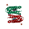
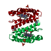
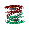
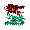
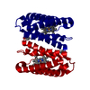
 Components Components | CYTOCHROME C' | |||||||||
 Keywords Keywords | ELECTRON TRANSPORT / HAEMOPROTEIN / 4-HELIX BUNDLE | |||||||||
| Function / homology |  Function and homology information Function and homology informationelectron transport chain / periplasmic space / electron transfer activity / iron ion binding / heme binding Similarity search - Function | |||||||||
| Biological species |  ACHROMOBACTER XYLOSOXIDANS (bacteria) ACHROMOBACTER XYLOSOXIDANS (bacteria) | |||||||||
| Method |  X-RAY DIFFRACTION / SYNCHROTRON / FOURIER SYNTHESIS / Resolution: 1.05 Å X-RAY DIFFRACTION / SYNCHROTRON / FOURIER SYNTHESIS / Resolution: 1.05 Å | |||||||||
 Authors Authors | Antonyuk, S.V. / Rustage, N. / Eady, R.R. / Hasnain, S.S. | |||||||||
 Citation Citation | Journal: Proc.Natl.Acad.Sci.USA / Year: 2011 Title: Carbon Monoxide Poisoning is Prevented by the Energy Costs of Conformational Changes in Gas- Binding Haemproteins. Authors: Antonyuk, S.V. / Rustage, N. / Petersen, C.A. / Arnst, J.L. / Heyes, D.J. / Sharma, R. / Berry, N.G. / Scrutton, N.S. / Eady, R.R. / Andrew, C.R. / Hasnain, S.S. | |||||||||
| History |
|
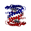
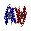
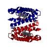
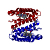
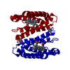
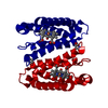
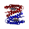
- Structure visualization
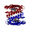
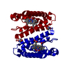
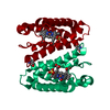
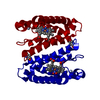
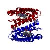
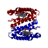
| Structure viewer | Molecule: MolmilJmol/JSmol |
|---|
- Downloads & links
-Download
| PDBx/mmCIF format | 3ztz.cif.gz | 88.1 KB | Display | PDBx/mmCIF format |
|---|---|---|---|---|
| PDB format | pdb3ztz.ent.gz | 67.3 KB | Display | PDB format |
| PDBx/mmJSON format | 3ztz.json.gz | Tree view | PDBx/mmJSON format | |
| Others |  Other downloads Other downloads |
-Validation report
| Arichive directory | https://data.pdbj.org/pub/pdb/validation_reports/zt/3ztzftp://data.pdbj.org/pub/pdb/validation_reports/zt/3ztz | HTTPS FTP |
|---|
-Related structure data
| Related structure data |  2ykzC  2yl0C 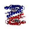 2yl1C 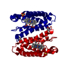 2yl3C 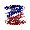 2yl7C 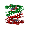 2yldC  2ylgSC  2yliC  3zqvC  3zqyC  3ztmC 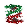 3zwiC C: citing same article ( S: Starting model for refinement |
|---|---|
| Similar structure data |


-Links
 PDBj
PDBj


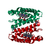
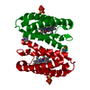
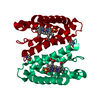
- Assembly
| Deposited unit | 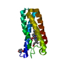
| ||||||||
|---|---|---|---|---|---|---|---|---|---|
| 1 | 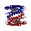
| ||||||||
| Unit cell |
|
-Components
| #1: Protein | Mass: 13575.336 Da / Num. of mol.: 1 / Mutation: YES Source method: isolated from a genetically manipulated source Source: (gene. exp.) ACHROMOBACTER XYLOSOXIDANS (bacteria) / Production host: | ||
|---|---|---|---|
| #2: Chemical | ChemComp-HEC /   Mass: 618.503 Da / Num. of mol.: 1 / Source method: obtained synthetically / Formula: C34H34FeN4O4 Mass: 618.503 Da / Num. of mol.: 1 / Source method: obtained synthetically / Formula: C34H34FeN4O4 | ||
| #3: Chemical | ChemComp-CMO /   Mass: 28.010 Da / Num. of mol.: 1 / Source method: obtained synthetically / Formula: CO Mass: 28.010 Da / Num. of mol.: 1 / Source method: obtained synthetically / Formula: CO | ||
| #4: Water | ChemComp-HOH /  Mass: 18.015 Da / Num. of mol.: 274 / Source method: isolated from a natural source / Formula: H2O Mass: 18.015 Da / Num. of mol.: 274 / Source method: isolated from a natural source / Formula: H2O | ||
| Compound details | ENGINEERED| Has protein modification | Y | |
-Experimental details
-Experiment
| Experiment | Method: X-RAY DIFFRACTION / Number of used crystals: 1 |
|---|
- Sample preparation
| Crystal | Density Matthews: 2.76 Å3/Da / Density % sol: 55.49 % / Description: NONE |
|---|---|
| Crystal grow | pH: 7.5 / Details: AMMONIUM SULPHATE, HEPES PH 7.5. |
-Data collection
| Diffraction | Mean temperature: 100 K |
|---|---|
| Diffraction source | Source: SYNCHROTRON / Site: Diamond  / Beamline: I02 / Wavelength: 0.9 / Beamline: I02 / Wavelength: 0.9 |
| Detector | Type: ADSC CCD / Detector: CCD / Date: Jun 4, 2009 / Details: MIRRORS |
| Radiation | Monochromator: SI111 / Protocol: SINGLE WAVELENGTH / Monochromatic (M) / Laue (L): M / Scattering type: x-ray |
| Radiation wavelength | Wavelength: 0.9 Å / Relative weight: 1 |
| Reflection | Resolution: 1.04→50 Å / Num. obs: 74665 / % possible obs: 99.5 % / Observed criterion σ(I): 0 / Redundancy: 8.5 % / Rmerge(I) obs: 0.083 / Net I/σ(I): 22 |
| Reflection shell | Resolution: 1.04→1.08 Å / Redundancy: 3.4 % / Rmerge(I) obs: 0.38 / Mean I/σ(I) obs: 2.7 / % possible all: 97.6 |
- Processing
| Software |
| ||||||||||||||||||||
|---|---|---|---|---|---|---|---|---|---|---|---|---|---|---|---|---|---|---|---|---|---|
| Refinement | Method to determine structure: FOURIER SYNTHESIS Starting model: PDB ENTRY 2YLG Resolution: 1.05→10 Å / Num. parameters: 13792 / Num. restraintsaints: 0 / Cross valid method: FREE R-VALUE / σ(F): 0 / Stereochemistry target values: ENGH AND HUBER Details: AT THE LAST RUN OF REFINEMENT R-FREE FLAGS WERE REMOVED AND DATA WERE REFINED AGAINST ALL DATA.
| ||||||||||||||||||||
| Solvent computation | Solvent model: MOEWS & KRETSINGER, J.MOL.BIOL.91(1973)201-228 | ||||||||||||||||||||
| Refine analyze | Num. disordered residues: 80 / Occupancy sum hydrogen: 877.54 / Occupancy sum non hydrogen: 1219.9 | ||||||||||||||||||||
| Refinement step | Cycle: LAST / Resolution: 1.05→10 Å
|
