 Movie
Movie Controller

[English] 日本語
 Yorodumi
- PDB-2xl8: REDUCED STRUCTURE OF R124F MUTANT OF CYTOCHROME C' FROM ALCALIGEN... -
+ Open data

- Basic information
| Entry | Database: PDB / ID: 2xl8 | |||||||||
|---|---|---|---|---|---|---|---|---|---|---|
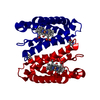
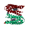
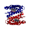
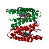
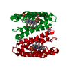
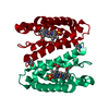
| Title | REDUCED STRUCTURE OF R124F MUTANT OF CYTOCHROME C' FROM ALCALIGENES XYLOSOXIDANS | |||||||||
 Components Components | CYTOCHROME C' | |||||||||
 Keywords Keywords | ELECTRON TRANSPORT / HAEMOPROTEIN / 4-HELIX BUNDLE | |||||||||
| Function / homology |  Function and homology information Function and homology informationelectron transport chain / periplasmic space / electron transfer activity / iron ion binding / heme binding Similarity search - Function | |||||||||
| Biological species |  ACHROMOBACTER XYLOSOXIDANS (bacteria) ACHROMOBACTER XYLOSOXIDANS (bacteria) | |||||||||
| Method |  X-RAY DIFFRACTION / SYNCHROTRON / MOLECULAR REPLACEMENT / Resolution: 1.14 Å X-RAY DIFFRACTION / SYNCHROTRON / MOLECULAR REPLACEMENT / Resolution: 1.14 Å | |||||||||
 Authors Authors | Hough, M.A. / Antonyuk, S.V. / Barbieri, S. / Rustage, N. / McKay, A.L. / Servid, A.E. / Eady, R.R. / Andrew, C.R. / Hasnain, S.S. | |||||||||
 Citation Citation | Journal: J.Mol.Biol. / Year: 2011 Title: Distal-to-Proximal No Conversion in Hemoproteins: The Role of the Proximal Pocket. Authors: Hough, M.A. / Antonyuk, S.V. / Barbieri, S. / Rustage, N. / Mckay, A.L. / Servid, A.E. / Eady, R.R. / Andrew, C.R. / Hasnain, S.S. | |||||||||
| History |
|
- Structure visualization
| Structure viewer | Molecule: MolmilJmol/JSmol |
|---|
- Downloads & links
-Download
| PDBx/mmCIF format | 2xl8.cif.gz | 85.7 KB | Display | PDBx/mmCIF format |
|---|---|---|---|---|
| PDB format | pdb2xl8.ent.gz | 65 KB | Display | PDB format |
| PDBx/mmJSON format | 2xl8.json.gz | Tree view | PDBx/mmJSON format | |
| Others |  Other downloads Other downloads |
-Validation report
| Arichive directory | https://data.pdbj.org/pub/pdb/validation_reports/xl/2xl8ftp://data.pdbj.org/pub/pdb/validation_reports/xl/2xl8 | HTTPS FTP |
|---|
-Related structure data
| Related structure data | 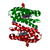 2xl6C 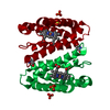 2xldC 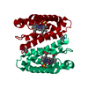 2xleC  2xlhC 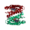 2xlmC 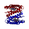 2xloC 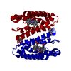 2xlvC 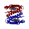 2xlwC 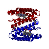 2xm0C  2xm4C  1e84S  2xl5 C: citing same article ( S: Starting model for refinement |
|---|---|
| Similar structure data |


-Links
 PDBj
PDBj


- Assembly
| Deposited unit | 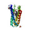
| ||||||||||||
|---|---|---|---|---|---|---|---|---|---|---|---|---|---|
| 1 | 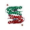
| ||||||||||||
| Unit cell |
| ||||||||||||
| Components on special symmetry positions |
|
-Components
| #1: Protein | Mass: 13621.423 Da / Num. of mol.: 1 / Mutation: YES Source method: isolated from a genetically manipulated source Source: (gene. exp.) ACHROMOBACTER XYLOSOXIDANS (bacteria) / Strain: NCIB 11015 / Plasmid: PTCP-1 / Production host: | ||
|---|---|---|---|
| #2: Chemical | ChemComp-HEC /   Mass: 618.503 Da / Num. of mol.: 1 / Source method: obtained synthetically / Formula: C34H34FeN4O4 Mass: 618.503 Da / Num. of mol.: 1 / Source method: obtained synthetically / Formula: C34H34FeN4O4 | ||
| #3: Chemical | ChemComp-SO4 /   Mass: 96.063 Da / Num. of mol.: 1 / Source method: obtained synthetically / Formula: SO4 Mass: 96.063 Da / Num. of mol.: 1 / Source method: obtained synthetically / Formula: SO4 | ||
| #4: Water | ChemComp-HOH /  Mass: 18.015 Da / Num. of mol.: 305 / Source method: isolated from a natural source / Formula: H2O Mass: 18.015 Da / Num. of mol.: 305 / Source method: isolated from a natural source / Formula: H2O | ||
| Compound details | ENGINEERED| Has protein modification | Y | |
-Experimental details
-Experiment
| Experiment | Method: X-RAY DIFFRACTION / Number of used crystals: 1 |
|---|
- Sample preparation
| Crystal | Density Matthews: 2.62 Å3/Da / Density % sol: 53 % / Description: NONE |
|---|---|
| Crystal grow | Temperature: 294 K / Method: vapor diffusion, hanging drop / pH: 7.5 / Details: SULPHATE AMMONIUM, TRIS PH 7.5 |
-Data collection
| Diffraction | Mean temperature: 100 K |
|---|---|
| Diffraction source | Source: SYNCHROTRON / Site: SRS  / Beamline: PX10.1 / Wavelength: 0.92 / Beamline: PX10.1 / Wavelength: 0.92 |
| Detector | Type: MARRESEARCH / Detector: CCD / Date: Mar 2, 2007 / Details: MIRRORS |
| Radiation | Monochromator: SI111 / Protocol: SINGLE WAVELENGTH / Monochromatic (M) / Laue (L): M / Scattering type: x-ray |
| Radiation wavelength | Wavelength: 0.92 Å / Relative weight: 1 |
| Reflection | Resolution: 1.14→23 Å / Num. obs: 56022 / % possible obs: 97.4 % / Observed criterion σ(I): 0 / Redundancy: 8.9 % / Biso Wilson estimate: 12.35 Å2 / Rmerge(I) obs: 0.056 / Net I/σ(I): 31 |
| Reflection shell | Resolution: 1.14→1.18 Å / Redundancy: 3.9 % / Rmerge(I) obs: 0.219 / Mean I/σ(I) obs: 5 / % possible all: 83.9 |
- Processing
| Software |
| ||||||||||||||||||||||||||||||||||||||||||||||||||||||||||||||||||||||||||||||||||||||||||||||||||||||||||||||||||||||||||||||||||||||||||||||||||||||||||||||||||||||||||||||||||||||
|---|---|---|---|---|---|---|---|---|---|---|---|---|---|---|---|---|---|---|---|---|---|---|---|---|---|---|---|---|---|---|---|---|---|---|---|---|---|---|---|---|---|---|---|---|---|---|---|---|---|---|---|---|---|---|---|---|---|---|---|---|---|---|---|---|---|---|---|---|---|---|---|---|---|---|---|---|---|---|---|---|---|---|---|---|---|---|---|---|---|---|---|---|---|---|---|---|---|---|---|---|---|---|---|---|---|---|---|---|---|---|---|---|---|---|---|---|---|---|---|---|---|---|---|---|---|---|---|---|---|---|---|---|---|---|---|---|---|---|---|---|---|---|---|---|---|---|---|---|---|---|---|---|---|---|---|---|---|---|---|---|---|---|---|---|---|---|---|---|---|---|---|---|---|---|---|---|---|---|---|---|---|---|---|
| Refinement | Method to determine structure: MOLECULAR REPLACEMENT Starting model: PDB ENTRY 1E84 Resolution: 1.14→46.47 Å / Cor.coef. Fo:Fc: 0.977 / Cor.coef. Fo:Fc free: 0.97 / SU B: 0.807 / SU ML: 0.017 / Cross valid method: THROUGHOUT / ESU R: 0.032 / ESU R Free: 0.032 / Stereochemistry target values: MAXIMUM LIKELIHOOD Details: HYDROGENS HAVE BEEN ADDED IN THE RIDING POSITIONS. U VALUES REFINED INDIVIDUALLY.
| ||||||||||||||||||||||||||||||||||||||||||||||||||||||||||||||||||||||||||||||||||||||||||||||||||||||||||||||||||||||||||||||||||||||||||||||||||||||||||||||||||||||||||||||||||||||
| Solvent computation | Ion probe radii: 0.8 Å / Shrinkage radii: 0.8 Å / VDW probe radii: 1.4 Å / Solvent model: MASK | ||||||||||||||||||||||||||||||||||||||||||||||||||||||||||||||||||||||||||||||||||||||||||||||||||||||||||||||||||||||||||||||||||||||||||||||||||||||||||||||||||||||||||||||||||||||
| Displacement parameters | Biso mean: 12.758 Å2
| ||||||||||||||||||||||||||||||||||||||||||||||||||||||||||||||||||||||||||||||||||||||||||||||||||||||||||||||||||||||||||||||||||||||||||||||||||||||||||||||||||||||||||||||||||||||
| Refinement step | Cycle: LAST / Resolution: 1.14→46.47 Å
| ||||||||||||||||||||||||||||||||||||||||||||||||||||||||||||||||||||||||||||||||||||||||||||||||||||||||||||||||||||||||||||||||||||||||||||||||||||||||||||||||||||||||||||||||||||||
| Refine LS restraints |
|