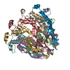
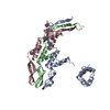
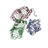
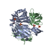
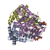
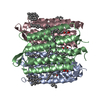
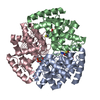
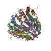
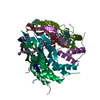
A: TERMINASE SMALL SUBUNIT B: TERMINASE SMALL SUBUNIT C: TERMINASE SMALL SUBUNIT D: TERMINASE SMALL SUBUNIT E: TERMINASE SMALL SUBUNIT F: TERMINASE SMALL SUBUNIT G: TERMINASE SMALL SUBUNIT H: TERMINASE SMALL SUBUNIT I: TERMINASE SMALL SUBUNIT J: TERMINASE SMALL SUBUNIT K: TERMINASE SMALL SUBUNIT L: TERMINASE SMALL SUBUNIT M: TERMINASE SMALL SUBUNIT N: TERMINASE SMALL SUBUNIT O: TERMINASE SMALL SUBUNIT P: TERMINASE SMALL SUBUNIT Q: TERMINASE SMALL SUBUNIT R: TERMINASE SMALL SUBUNIT hetero molecules
A: TERMINASE SMALL SUBUNIT B: TERMINASE SMALL SUBUNIT C: TERMINASE SMALL SUBUNIT D: TERMINASE SMALL SUBUNIT E: TERMINASE SMALL SUBUNIT F: TERMINASE SMALL SUBUNIT G: TERMINASE SMALL SUBUNIT H: TERMINASE SMALL SUBUNIT I: TERMINASE SMALL SUBUNIT
J: TERMINASE SMALL SUBUNIT K: TERMINASE SMALL SUBUNIT L: TERMINASE SMALL SUBUNIT M: TERMINASE SMALL SUBUNIT N: TERMINASE SMALL SUBUNIT O: TERMINASE SMALL SUBUNIT P: TERMINASE SMALL SUBUNIT Q: TERMINASE SMALL SUBUNIT R: TERMINASE SMALL SUBUNIT hetero molecules
Resolution: 1.68→22.1 Å / Cor.coef. Fo:Fc: 0.938 / Cor.coef. Fo:Fc free: 0.927 / SU B: 6.191 / SU ML: 0.082 / Cross valid method: THROUGHOUT / ESU R: 0.029 / ESU R Free: 0.023 / Stereochemistry target values: MAXIMUM LIKELIHOOD Details: HYDROGENS HAVE BEEN ADDED IN THE RIDING POSITIONS. U VALUES REFINED INDIVIDUALLY.
Rfactor
Num. reflection
% reflection
Selection details
Rfree
0.23357
1231
1 %
RANDOM
Rwork
0.18289
-
-
-
obs
0.18339
126317
83.01 %
-
Solvent computation
Ion probe radii: 0.8 Å / Shrinkage radii: 0.8 Å / VDW probe radii: 1.2 Å / Solvent model: MASK
 Movie
Movie Controller

 Yorodumi
 Open data
 Basic information
 Components
 Keywords
 Function and homology information
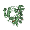
 BACILLUS PHAGE SF6 (virus)
 X-RAY DIFFRACTION /
  Authors
 Citation
 Structure visualization
 Downloads & links
 Other downloads
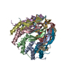


 PDBj
PDBj Assembly


 Mass: 39.098 Da / Num. of mol.: 2 / Source method: obtained synthetically / Formula: K
 Mass: 18.015 Da / Num. of mol.: 604 / Source method: isolated from a natural source / Formula: H2O
 Sample preparation
 / Beamline: BM14 / Wavelength: 0.97
  Processing