 Movie
Movie Controller

[English] 日本語
 Yorodumi
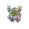
- PDB-3zqq: Crystal structure of the full-length small terminase from a SPP1-... -
+ Open data

- Basic information
| Entry | Database: PDB / ID: 3zqq | ||||||
|---|---|---|---|---|---|---|---|
| Title | Crystal structure of the full-length small terminase from a SPP1-like bacteriophage | ||||||
 Components Components | TERMINASE SMALL SUBUNIT | ||||||
 Keywords Keywords | DNA BINDING PROTEIN / DNA-BINDING PROTEIN / DNA PACKAGING | ||||||
| Function / homology |  Function and homology information Function and homology information | ||||||
| Biological species |  BACILLUS PHAGE SF6 (virus) BACILLUS PHAGE SF6 (virus) | ||||||
| Method |  X-RAY DIFFRACTION / SYNCHROTRON / MOLECULAR REPLACEMENT / Resolution: 4 Å X-RAY DIFFRACTION / SYNCHROTRON / MOLECULAR REPLACEMENT / Resolution: 4 Å | ||||||
 Authors Authors | Buttner, C.R. / Chechik, M. / Ortiz-Lombardia, M. / Smits, C. / Chechik, V. / Jeschke, G. / Dykeman, E. / Benini, S. / Alonso, J.C. / Antson, A.A. | ||||||
 Citation Citation | Journal: Proc.Natl.Acad.Sci.USA / Year: 2012 Title: Structural Basis for DNA Recognition and Loading Into a Viral Packaging Motor. Authors: Buttner, C.R. / Chechik, M. / Ortiz-Lombardia, M. / Smits, C. / Ebong, I.O. / Chechik, V. / Jeschke, G. / Dykeman, E. / Benini, S. / Robinson, C.V. / Alonso, J.C. / Antson, A.A. | ||||||
| History |
| ||||||
| Remark 700 | SHEET DETERMINATION METHOD: AUTHOR PROVIDED. |
- Structure visualization
| Structure viewer | Molecule: MolmilJmol/JSmol |
|---|
- Downloads & links
-Download
| PDBx/mmCIF format | 3zqq.cif.gz | 73 KB | Display | PDBx/mmCIF format |
|---|---|---|---|---|
| PDB format | pdb3zqq.ent.gz | 55.6 KB | Display | PDB format |
| PDBx/mmJSON format | 3zqq.json.gz | Tree view | PDBx/mmJSON format | |
| Others |  Other downloads Other downloads |
-Validation report
| Arichive directory | https://data.pdbj.org/pub/pdb/validation_reports/zq/3zqqftp://data.pdbj.org/pub/pdb/validation_reports/zq/3zqq | HTTPS FTP |
|---|
-Related structure data
| Related structure data | 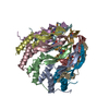 3zqmC 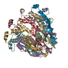 3zqnC 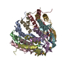 3zqoC  3zqpSC  2cmpS C: citing same article ( S: Starting model for refinement |
|---|---|
| Similar structure data |


-Links
 PDBj
PDBj- Assembly
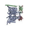
| Deposited unit | 
| ||||||||
|---|---|---|---|---|---|---|---|---|---|
| 1 | 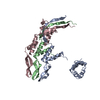
| ||||||||
| Unit cell |
|
-Components
| #1: Protein | Mass: 18069.576 Da / Num. of mol.: 3 Source method: isolated from a genetically manipulated source Source: (gene. exp.) BACILLUS PHAGE SF6 (virus) / Plasmid: PET28A / Production host:  |
|---|
-Experimental details
-Experiment
| Experiment | Method: X-RAY DIFFRACTION / Number of used crystals: 1 |
|---|
- Sample preparation
| Crystal | Density Matthews: 3.07 Å3/Da / Density % sol: 60 % Description: MR WAS PERFORMED USING THREE NEIGHBORING SUBUNITS |
|---|---|
| Crystal grow | Details: 0.2 M MGCL2, 8 % PEG 2000, 8 % PEG 550MME, 0.1 M TRIS PH 8.0 |
-Data collection
| Diffraction | Mean temperature: 100 K |
|---|---|
| Diffraction source | Source: SYNCHROTRON / Site: APS  / Beamline: 14-ID-B / Wavelength: 0.934 / Beamline: 14-ID-B / Wavelength: 0.934 |
| Detector | Type: MARRESEARCH / Detector: CCD / Date: Feb 4, 2004 |
| Radiation | Protocol: SINGLE WAVELENGTH / Monochromatic (M) / Laue (L): M / Scattering type: x-ray |
| Radiation wavelength | Wavelength: 0.934 Å / Relative weight: 1 |
| Reflection | Resolution: 4.01→107 Å / Num. obs: 3308 / % possible obs: 62 % / Observed criterion σ(I): 2 / Redundancy: 8.5 % / Rmerge(I) obs: 0.08 / Net I/σ(I): 6.6 |
| Reflection shell | Resolution: 4.01→4.23 Å / Redundancy: 8.6 % / Rmerge(I) obs: 0.19 / Mean I/σ(I) obs: 4.1 / % possible all: 18 |
- Processing
| Software |
| ||||||||||||||||||||||||||||||||||||||||||||||||||||||||||||
|---|---|---|---|---|---|---|---|---|---|---|---|---|---|---|---|---|---|---|---|---|---|---|---|---|---|---|---|---|---|---|---|---|---|---|---|---|---|---|---|---|---|---|---|---|---|---|---|---|---|---|---|---|---|---|---|---|---|---|---|---|---|
| Refinement | Method to determine structure: MOLECULAR REPLACEMENT Starting model: PDB ENTRIES 2CMP AND 3ZQP Resolution: 4→107 Å / Data cutoff high absF: 10000 / Cross valid method: THROUGHOUT / σ(F): 0 Details: THE THIRD DNA-BINDING DOMAIN (DBD OF CHAIN B) IS NOT SUFFICIENTLY DEFINED IN THE ELECTRON DENSITY AND WAS NOT MODELED.
| ||||||||||||||||||||||||||||||||||||||||||||||||||||||||||||
| Solvent computation | Bsol: 207.145 Å2 / ksol: 0.37 e/Å3 | ||||||||||||||||||||||||||||||||||||||||||||||||||||||||||||
| Displacement parameters | Biso mean: 220 Å2
| ||||||||||||||||||||||||||||||||||||||||||||||||||||||||||||
| Refinement step | Cycle: LAST / Resolution: 4→107 Å
| ||||||||||||||||||||||||||||||||||||||||||||||||||||||||||||
| Refine LS restraints |
| ||||||||||||||||||||||||||||||||||||||||||||||||||||||||||||
| LS refinement shell | Resolution: 4.01→4.23 Å / Rfactor Rfree: 0.2011 / Rfactor Rwork: 0.2026 / Total num. of bins used: 10 | ||||||||||||||||||||||||||||||||||||||||||||||||||||||||||||
| Xplor file |
|
