 Movie
Movie Controller


+ Open data

- Basic information
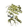
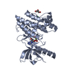
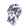
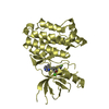
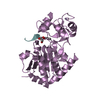
| Entry | Database: PDB / ID: 3zlc | ||||||
|---|---|---|---|---|---|---|---|
| Title | Crystal Structure of Erv41p | ||||||
 Components Components | ER-DERIVED VESICLES PROTEIN ERV41 | ||||||
 Keywords Keywords | PROTEIN TRANSPORT / EARLY SECRETORY PATHWAY / ENDOPLASMIC RETICULUM / COPII VESICLE | ||||||
| Function / homology |  Function and homology information Function and homology informationretrograde cargo receptor complex, Golgi to ER / COPII-coated ER to Golgi transport vesicle / retrograde vesicle-mediated transport, Golgi to endoplasmic reticulum / endoplasmic reticulum to Golgi vesicle-mediated transport / ER to Golgi transport vesicle membrane / protein transport / Golgi membrane / endoplasmic reticulum membrane / endoplasmic reticulum / identical protein binding Similarity search - Function | ||||||
| Biological species |  | ||||||
| Method |  X-RAY DIFFRACTION / SYNCHROTRON / MOLECULAR REPLACEMENT / Resolution: 1.999 Å X-RAY DIFFRACTION / SYNCHROTRON / MOLECULAR REPLACEMENT / Resolution: 1.999 Å | ||||||
 Authors Authors | Biterova, E.I. / Svard, M. / Possner, D.D.D. / Guy, J.E. | ||||||
 Citation Citation | Journal: J.Mol.Biol. / Year: 2013 Title: The Crystal Structure of the Lumenal Domain of Erv41P, a Protein Involved in Transport between the Endoplasmic Reticulum and Golgi Apparatus Authors: Biterova, E.I. / Svard, M. / Possner, D.D.D. / Guy, J.E. | ||||||
| History |
|
- Structure visualization
| Structure viewer | Molecule: MolmilJmol/JSmol |
|---|
- Downloads & links
-Download
| PDBx/mmCIF format | 3zlc.cif.gz | 196.5 KB | Display | PDBx/mmCIF format |
|---|---|---|---|---|
| PDB format | pdb3zlc.ent.gz | 158.4 KB | Display | PDB format |
| PDBx/mmJSON format | 3zlc.json.gz | Tree view | PDBx/mmJSON format | |
| Others |  Other downloads Other downloads |
-Validation report
| Arichive directory | https://data.pdbj.org/pub/pdb/validation_reports/zl/3zlcftp://data.pdbj.org/pub/pdb/validation_reports/zl/3zlc | HTTPS FTP |
|---|
-Related structure data
| Similar structure data |
|---|


-Links
 PDBj
PDBj- Assembly
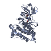
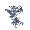
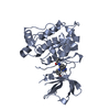
| Deposited unit | 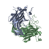
| ||||||||
|---|---|---|---|---|---|---|---|---|---|
| 1 | 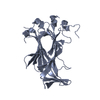
| ||||||||
| 2 | 
| ||||||||
| 3 |
| ||||||||
| Unit cell |
| ||||||||
| Noncrystallographic symmetry (NCS) | NCS oper: (Code: given Matrix: (0.8662, 0.0747, 0.494), Vector: |
-Components
| #1: Protein | Mass: 29109.650 Da / Num. of mol.: 2 Source method: isolated from a genetically manipulated source Source: (gene. exp.) Strain: S288C / Plasmid: PFHMSP-N / Cell line (production host): SF9 / Production host:   SPODOPTERA FRUGIPERDA (fall armyworm) / References: UniProt: Q04651 SPODOPTERA FRUGIPERDA (fall armyworm) / References: UniProt: Q04651#2: Water | ChemComp-HOH / |  Mass: 18.015 Da / Num. of mol.: 240 / Source method: isolated from a natural source / Formula: H2O Mass: 18.015 Da / Num. of mol.: 240 / Source method: isolated from a natural source / Formula: H2OHas protein modification | Y | |
|---|
-Experimental details
-Experiment
| Experiment | Method: X-RAY DIFFRACTION / Number of used crystals: 1 |
|---|
- Sample preparation
| Crystal | Density Matthews: 2.07 Å3/Da / Density % sol: 40 % / Description: NONE |
|---|---|
| Crystal grow | Details: 0.2 M NH4NO3, 16 % PEG 3350 |
-Data collection
| Diffraction | Mean temperature: 100 K |
|---|---|
| Diffraction source | Source: SYNCHROTRON / Site: ESRF  / Beamline: ID14-1 / Wavelength: 0.934 / Beamline: ID14-1 / Wavelength: 0.934 |
| Detector | Type: ADSC QUANTUM 210 / Detector: CCD / Date: Jun 25, 2012 |
| Radiation | Protocol: SINGLE WAVELENGTH / Monochromatic (M) / Laue (L): M / Scattering type: x-ray |
| Radiation wavelength | Wavelength: 0.934 Å / Relative weight: 1 |
| Reflection | Resolution: 2→48.14 Å / Num. obs: 31843 / % possible obs: 98.8 % / Observed criterion σ(I): 2 / Redundancy: 3.9 % / Biso Wilson estimate: 23.16 Å2 / Rmerge(I) obs: 0.05 / Net I/σ(I): 19.41 |
| Reflection shell | Resolution: 2→2.1 Å / Redundancy: 3.2 % / Rmerge(I) obs: 0.27 / Mean I/σ(I) obs: 4.41 / % possible all: 92.2 |
- Processing
| Software |
| |||||||||||||||||||||||||||||||||||||||||||||||||||||||||||||||||||||||||||||||||||||||||||||||||||||||||
|---|---|---|---|---|---|---|---|---|---|---|---|---|---|---|---|---|---|---|---|---|---|---|---|---|---|---|---|---|---|---|---|---|---|---|---|---|---|---|---|---|---|---|---|---|---|---|---|---|---|---|---|---|---|---|---|---|---|---|---|---|---|---|---|---|---|---|---|---|---|---|---|---|---|---|---|---|---|---|---|---|---|---|---|---|---|---|---|---|---|---|---|---|---|---|---|---|---|---|---|---|---|---|---|---|---|---|
| Refinement | Method to determine structure: MOLECULAR REPLACEMENT / Resolution: 1.999→48.14 Å / SU ML: 0.16 / σ(F): 2 / Phase error: 19.75 / Stereochemistry target values: ML
| |||||||||||||||||||||||||||||||||||||||||||||||||||||||||||||||||||||||||||||||||||||||||||||||||||||||||
| Solvent computation | Shrinkage radii: 0.9 Å / VDW probe radii: 1.11 Å / Solvent model: FLAT BULK SOLVENT MODEL | |||||||||||||||||||||||||||||||||||||||||||||||||||||||||||||||||||||||||||||||||||||||||||||||||||||||||
| Displacement parameters | Biso mean: 36.4 Å2 | |||||||||||||||||||||||||||||||||||||||||||||||||||||||||||||||||||||||||||||||||||||||||||||||||||||||||
| Refinement step | Cycle: LAST / Resolution: 1.999→48.14 Å
| |||||||||||||||||||||||||||||||||||||||||||||||||||||||||||||||||||||||||||||||||||||||||||||||||||||||||
| Refine LS restraints |
| |||||||||||||||||||||||||||||||||||||||||||||||||||||||||||||||||||||||||||||||||||||||||||||||||||||||||
| LS refinement shell |
| |||||||||||||||||||||||||||||||||||||||||||||||||||||||||||||||||||||||||||||||||||||||||||||||||||||||||
| Refinement TLS params. | Method: refined / Refine-ID: X-RAY DIFFRACTION
| |||||||||||||||||||||||||||||||||||||||||||||||||||||||||||||||||||||||||||||||||||||||||||||||||||||||||
| Refinement TLS group |
|
