 Movie
Movie Controller


+ Open data

- Basic information
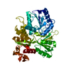
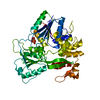
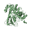
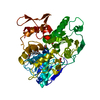
| Entry | Database: PDB / ID: 3zl7 | ||||||
|---|---|---|---|---|---|---|---|
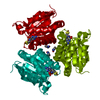
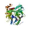
| Title | BACE2 FYNOMER COMPLEX | ||||||
 Components Components |
| ||||||
 Keywords Keywords | HYDROLASE / ASPARTYL PROTEASE | ||||||
| Function / homology |  Function and homology information Function and homology informationmemapsin 1 / negative regulation of amyloid precursor protein biosynthetic process / melanosome membrane / melanosome organization / peptide hormone processing / membrane protein ectodomain proteolysis / amyloid-beta metabolic process / trans-Golgi network / protein processing / glucose homeostasis ...memapsin 1 / negative regulation of amyloid precursor protein biosynthetic process / melanosome membrane / melanosome organization / peptide hormone processing / membrane protein ectodomain proteolysis / amyloid-beta metabolic process / trans-Golgi network / protein processing / glucose homeostasis / aspartic-type endopeptidase activity / endosome / Golgi apparatus / endoplasmic reticulum / proteolysis / membrane / plasma membrane Similarity search - Function | ||||||
| Biological species |  HOMO SAPIENS (human) HOMO SAPIENS (human)SYNTHETIC CONSTRUCT (others) | ||||||
| Method |  X-RAY DIFFRACTION / SYNCHROTRON / MOLECULAR REPLACEMENT / Resolution: 3.2 Å X-RAY DIFFRACTION / SYNCHROTRON / MOLECULAR REPLACEMENT / Resolution: 3.2 Å | ||||||
 Authors Authors | Banner, D.W. / Kuglstatter, A. / Benz, J. / Stihle, M. / Ruf, A. | ||||||
 Citation Citation | Journal: Acta Crystallogr.,Sect.D / Year: 2013 Title: Mapping the Conformational Space Accessible to Bace2 Using Surface Mutants and Co-Crystals with Fab-Fragments, Fynomers, and Xaperones Authors: Banner, D.W. / Gsell, B. / Benz, J. / Bertschinger, J. / Burger, D. / Brack, S. / Cuppuleri, S. / Debulpaep, M. / Gast, A. / Grabulovski, D. / Hennig, M. / Hilpert, H. / Huber, W. / ...Authors: Banner, D.W. / Gsell, B. / Benz, J. / Bertschinger, J. / Burger, D. / Brack, S. / Cuppuleri, S. / Debulpaep, M. / Gast, A. / Grabulovski, D. / Hennig, M. / Hilpert, H. / Huber, W. / Kuglstatter, A. / Kusznir, E. / Laeremans, T. / Matile, H. / Miscenic, C. / Rufer, A. / Schlatter, D. / Steyeart, J. / Stihle, M. / Thoma, R. / Weber, M. / Ruf, A. | ||||||
| History |
| ||||||
| Remark 700 | SHEET DETERMINATION METHOD: DSSP THE SHEETS PRESENTED AS "AB" IN EACH CHAIN ON SHEET RECORDS BELOW ... SHEET DETERMINATION METHOD: DSSP THE SHEETS PRESENTED AS "AB" IN EACH CHAIN ON SHEET RECORDS BELOW IS ACTUALLY AN 6-STRANDED BARREL THIS IS REPRESENTED BY A 7-STRANDED SHEET IN WHICH THE FIRST AND LAST STRANDS ARE IDENTICAL. |
- Structure visualization
| Structure viewer | Molecule: MolmilJmol/JSmol |
|---|
- Downloads & links
-Download
| PDBx/mmCIF format | 3zl7.cif.gz | 98.4 KB | Display | PDBx/mmCIF format |
|---|---|---|---|---|
| PDB format | pdb3zl7.ent.gz | 74 KB | Display | PDB format |
| PDBx/mmJSON format | 3zl7.json.gz | Tree view | PDBx/mmJSON format | |
| Others |  Other downloads Other downloads |
-Validation report
| Arichive directory | https://data.pdbj.org/pub/pdb/validation_reports/zl/3zl7ftp://data.pdbj.org/pub/pdb/validation_reports/zl/3zl7 | HTTPS FTP |
|---|
-Related structure data
| Related structure data | 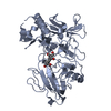 3zkgC  3zkiC  3zkmC  3zknC  3zkqSC  3zksC  3zkxC  3zovC 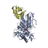 4belC  4bfbC  4ag1S C: citing same article ( S: Starting model for refinement |
|---|---|
| Similar structure data |


-Links
 PDBj
PDBj

- Assembly
| Deposited unit | 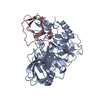
| ||||||||
|---|---|---|---|---|---|---|---|---|---|
| 1 |
| ||||||||
| Unit cell |
|
-Components
| #1: Protein | Mass: 42047.355 Da / Num. of mol.: 1 / Fragment: EXTRACELLULAR DOMAIN, RESIDUES 75-460 / Mutation: YES Source method: isolated from a genetically manipulated source Source: (gene. exp.) HOMO SAPIENS (human) / Production host:  |
|---|---|
| #2: Protein | Mass: 10325.441 Da / Num. of mol.: 1 Source method: isolated from a genetically manipulated source Source: (gene. exp.) SYNTHETIC CONSTRUCT (others) / Production host: |
| #3: Water | ChemComp-HOH /  Mass: 18.015 Da / Num. of mol.: 22 / Source method: isolated from a natural source / Formula: H2O Mass: 18.015 Da / Num. of mol.: 22 / Source method: isolated from a natural source / Formula: H2O |
| Has protein modification | Y |
| Sequence details | THE PDB FILE IS NUMBERED AFTER PDB-ENTRY 2EWY WHICH HAS NUMBERS 62 LESS THAN THE DATA BANK SEQUENCE. ...THE PDB FILE IS NUMBERED AFTER PDB-ENTRY 2EWY WHICH HAS NUMBERS 62 LESS THAN THE DATA BANK SEQUENCE. THE MUTATION HERE IS E269A IN THE PDB FILE AND E331A IN THE DATA BANK SEQUENCE. MODIFIED VERSION OF HUMAN FYN TYROSINE KINASE SH3 DOMAIN |
-Experimental details
-Experiment
| Experiment | Method: X-RAY DIFFRACTION / Number of used crystals: 1 |
|---|
- Sample preparation
| Crystal | Density Matthews: 2.46 Å3/Da / Density % sol: 50.1 % Description: DATA COMPROMISED BY DIFFUSE ICE RING AT 3.68A AND APERTURE SCATTER AT 3.24A |
|---|---|
| Crystal grow | pH: 5 / Details: 1.8M NA/K PHOSPHATE, PH 5.0 |
-Data collection
| Diffraction | Mean temperature: 100 K |
|---|---|
| Diffraction source | Source: SYNCHROTRON / Site: SLS  / Beamline: X10SA / Wavelength: 1 / Beamline: X10SA / Wavelength: 1 |
| Detector | Type: DECTRIS PILATUS 6M / Detector: PIXEL / Date: Mar 17, 2011 |
| Radiation | Protocol: SINGLE WAVELENGTH / Monochromatic (M) / Laue (L): M / Scattering type: x-ray |
| Radiation wavelength | Wavelength: 1 Å / Relative weight: 1 |
| Reflection | Resolution: 2.91→43.77 Å / Num. obs: 10238 / % possible obs: 94.5 % / Observed criterion σ(I): -3 / Redundancy: 11.6 % / Biso Wilson estimate: 91.84 Å2 / Rmerge(I) obs: 0.13 / Net I/σ(I): 12.9 |
| Reflection shell | Resolution: 2.91→3.01 Å / Redundancy: 12.7 % / Rmerge(I) obs: 0.74 / Mean I/σ(I) obs: 1.9 / % possible all: 100 |
- Processing
| Software |
| ||||||||||||||||||||||||||||||||||||||||||||||||||||||||||||||||||||||||||||||||||||||||||||||||||||||||||||||||||
|---|---|---|---|---|---|---|---|---|---|---|---|---|---|---|---|---|---|---|---|---|---|---|---|---|---|---|---|---|---|---|---|---|---|---|---|---|---|---|---|---|---|---|---|---|---|---|---|---|---|---|---|---|---|---|---|---|---|---|---|---|---|---|---|---|---|---|---|---|---|---|---|---|---|---|---|---|---|---|---|---|---|---|---|---|---|---|---|---|---|---|---|---|---|---|---|---|---|---|---|---|---|---|---|---|---|---|---|---|---|---|---|---|---|---|---|
| Refinement | Method to determine structure: MOLECULAR REPLACEMENT Starting model: PDB ENTRIES 3ZKQ, 4AG1 Resolution: 3.2→43.77 Å / Cor.coef. Fo:Fc: 0.8572 / Cor.coef. Fo:Fc free: 0.7992 / Cross valid method: THROUGHOUT / σ(F): 0 / SU Rfree Blow DPI: 0.667 Details: IDEAL-DIST CONTACT TERM CONTACT SETUP. ALL ATOMS HAVE CCP4 ATOM TYPE FROM LIBRARY. ALL LOWER SYMMETRY SPACE GROUPS WERE TESTED AND REJECTED AS REFINEMENT STATISTICS NOT SIGNIFICANTLY BETTER
| ||||||||||||||||||||||||||||||||||||||||||||||||||||||||||||||||||||||||||||||||||||||||||||||||||||||||||||||||||
| Displacement parameters | Biso mean: 80.23 Å2
| ||||||||||||||||||||||||||||||||||||||||||||||||||||||||||||||||||||||||||||||||||||||||||||||||||||||||||||||||||
| Refine analyze | Luzzati coordinate error obs: 0.86 Å | ||||||||||||||||||||||||||||||||||||||||||||||||||||||||||||||||||||||||||||||||||||||||||||||||||||||||||||||||||
| Refinement step | Cycle: LAST / Resolution: 3.2→43.77 Å
| ||||||||||||||||||||||||||||||||||||||||||||||||||||||||||||||||||||||||||||||||||||||||||||||||||||||||||||||||||
| Refine LS restraints |
| ||||||||||||||||||||||||||||||||||||||||||||||||||||||||||||||||||||||||||||||||||||||||||||||||||||||||||||||||||
| LS refinement shell | Resolution: 3.2→3.58 Å / Total num. of bins used: 5
|
