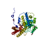
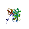
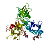
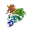
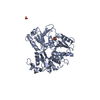
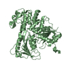
[English] 日本語
Yorodumi
- PDB-2bhr: Dengue virus RNA helicase -

+
Open data


ID or keywords:

Loading...

-
Basic information

Entry
Database: PDB / ID: 2bhr
TitleDengue virus RNA helicase
ComponentsRNA HELICASEHelicase
KeywordsHYDROLASE / HELICASE / NUCLEOSIDE TRIPHOSPHATASE / RNA-REPLICATION
Function / homology
Function and homology information


symbiont-mediated suppression of host JAK-STAT cascade via inhibition of host TYK2 activity / host cell mitochondrion / symbiont-mediated suppression of host JAK-STAT cascade via inhibition of STAT2 activity / symbiont-mediated suppression of host cytoplasmic pattern recognition receptor signaling pathway via inhibition of MAVS activity / ribonucleoside triphosphate phosphatase activity / viral capsid / : / double-stranded RNA binding / protein complex oligomerization / monoatomic ion channel activity ...symbiont-mediated suppression of host JAK-STAT cascade via inhibition of host TYK2 activity / host cell mitochondrion / symbiont-mediated suppression of host JAK-STAT cascade via inhibition of STAT2 activity / symbiont-mediated suppression of host cytoplasmic pattern recognition receptor signaling pathway via inhibition of MAVS activity / ribonucleoside triphosphate phosphatase activity / viral capsid / : / double-stranded RNA binding / protein complex oligomerization / monoatomic ion channel activity / clathrin-dependent endocytosis of virus by host cell / mRNA (nucleoside-2'-O-)-methyltransferase activity / mRNA 5'-cap (guanine-N7-)-methyltransferase activity / RNA helicase activity / host cell endoplasmic reticulum membrane / protein dimerization activity / induction by virus of host autophagy / viral RNA genome replication / RNA-dependent RNA polymerase activity / serine-type endopeptidase activity / fusion of virus membrane with host endosome membrane / viral envelope / symbiont-mediated suppression of host type I interferon-mediated signaling pathway / virion attachment to host cell / virion membrane / structural molecule activity / proteolysis / extracellular region / ATP binding / membrane / metal ion binding
Similarity search - Function
: / Flavivirus envelope glycoprotein E, stem/anchor domain / RNA-directed RNA polymerase, thumb domain, Flavivirus / Flavivirus RNA-directed RNA polymerase, thumb domain / Flavivirus capsid protein C superfamily / : / Flavivirus non-structural protein NS2B / Flavivirus NS3 helicase, C-terminal helical domain / Genome polyprotein, Flavivirus / Flavivirus non-structural protein NS4A ...: / Flavivirus envelope glycoprotein E, stem/anchor domain / RNA-directed RNA polymerase, thumb domain, Flavivirus / Flavivirus RNA-directed RNA polymerase, thumb domain / Flavivirus capsid protein C superfamily / : / Flavivirus non-structural protein NS2B / Flavivirus NS3 helicase, C-terminal helical domain / Genome polyprotein, Flavivirus / Flavivirus non-structural protein NS4A / Flavivirus non-structural protein NS2B / Flavivirus capsid protein C / Flavivirus non-structural protein NS4B / mRNA cap 0/1 methyltransferase / Flavivirus capsid protein C / Flavivirus non-structural protein NS4B / Flavivirus non-structural protein NS4A / Flavivirus NS2B domain profile. / mRNA cap 0 and cap 1 methyltransferase (EC 2.1.1.56 and EC 2.1.1.57) domain profile. / Flavivirus non-structural protein NS2A / Flavivirus non-structural protein NS2A / Flavivirus NS3, petidase S7 / Peptidase S7, Flavivirus NS3 serine protease / Flavivirus NS3 protease (NS3pro) domain profile. / Envelope glycoprotein M, flavivirus / Flavivirus envelope glycoprotein M / RNA-directed RNA polymerase, flavivirus / Flavivirus RNA-directed RNA polymerase, fingers and palm domains / Flavivirus non-structural Protein NS1 / Flavivirus non-structural protein NS1 / Envelope glycoprotein M superfamily, flavivirus / Flavivirus polyprotein propeptide / Flavivirus polyprotein propeptide superfamily / Flavivirus polyprotein propeptide / Flaviviral glycoprotein E, central domain, subdomain 1 / Flaviviral glycoprotein E, central domain, subdomain 2 / Flavivirus envelope glycoprotein E, Stem/Anchor domain / Flavivirus glycoprotein E, immunoglobulin-like domain / Flavivirus envelope glycoprotein E, Stem/Anchor domain superfamily / Flavivirus glycoprotein, immunoglobulin-like domain / Flavivirus glycoprotein central and dimerisation domain / Flavivirus glycoprotein, central and dimerisation domains / Ribosomal RNA methyltransferase, FtsJ domain / FtsJ-like methyltransferase / Flavivirus/Alphavirus glycoprotein, immunoglobulin-like domain superfamily / Flavivirus glycoprotein, central and dimerisation domain superfamily / Flaviviral glycoprotein E, dimerisation domain / DEAD box, Flavivirus / Flavivirus DEAD domain / helicase superfamily c-terminal domain / Immunoglobulin E-set / Superfamilies 1 and 2 helicase C-terminal domain profile. / Superfamilies 1 and 2 helicase ATP-binding type-1 domain profile. / DEAD-like helicases superfamily / Helicase, C-terminal / Helicase superfamily 1/2, ATP-binding domain / P-loop containing nucleotide triphosphate hydrolases / RNA-directed RNA polymerase, catalytic domain / RdRp of positive ssRNA viruses catalytic domain profile. / S-adenosyl-L-methionine-dependent methyltransferase superfamily / Peptidase S1, PA clan, chymotrypsin-like fold / Peptidase S1, PA clan / DNA/RNA polymerase superfamily / P-loop containing nucleoside triphosphate hydrolase / Rossmann fold / 3-Layer(aba) Sandwich / Alpha Beta
Similarity search - Domain/homology
Biological speciesDENGUE VIRUS 2
MethodX-RAY DIFFRACTION / SYNCHROTRON / MAD / Resolution: 2.8 Å
AuthorsXu, T. / Sampath, A. / Chao, A. / Wen, D. / Nanao, M. / Chene, P. / Vasudevan, S.G. / Lescar, J.
CitationJournal: J.Virol. / Year: 2005
Title: Structure of the Dengue Virus Helicase/Nucleoside Triphosphatase Catalytic Domain at a Resolution of 2.4 A.
Authors: Xu, T. / Sampath, A. / Chao, A. / Wen, D. / Nanao, M. / Chene, P. / Vasudevan, S.G. / Lescar, J.
History
DepositionJan 17, 2005Deposition site: PDBE / Processing site: PDBE
Revision 1.0Aug 3, 2005Provider: repository / Type: Initial release
Revision 1.1Jul 13, 2011Group: Source and taxonomy / Version format compliance

-
Structure visualization

Structure viewerMolecule:
MolmilJmol/JSmol

Downloads & links

-
Assembly

Deposited unit
A: RNA HELICASE
B: RNA HELICASE
hetero molecules


Theoretical massNumber of molelcules
Total (without water)103,4424
Polymers103,2502
Non-polymers1922
Water2,540141
1
A: RNA HELICASE
hetero molecules


Theoretical massNumber of molelcules
Total (without water)51,8173
Polymers51,6251
Non-polymers1922
Water181
TypeNameSymmetry operationNumber
identity operation1_555x,y,z1
MethodPQS
2
B: RNA HELICASE


Theoretical massNumber of molelcules
Total (without water)51,6251
Polymers51,6251
Non-polymers00
Water181
TypeNameSymmetry operationNumber
identity operation1_555x,y,z1
MethodPQS
Unit cell
Length a, b, c (Å)54.720, 165.970, 54.710
Angle α, β, γ (deg.)90.00, 101.88, 90.00
Int Tables number4
Space group name H-MP1211
Noncrystallographic symmetry (NCS)NCS oper: (Code: given
Matrix: (0.18936, -0.00077, -0.98191), (-0.1661, 0.98556, -0.03281), (0.96776, 0.16931, 0.1865))

-
Components

#1: Protein RNA HELICASE / Helicase


Mass: 51625.031 Da / Num. of mol.: 2 / Fragment: RESIDUES 1646-2093
Source method: isolated from a genetically manipulated source
Source: (gene. exp.) DENGUE VIRUS 2 / Description: AY037116, NUCLEOTIDES 5032-6301 / Production host: ESCHERICHIA COLI (E. coli) / Strain (production host): BL21 / Variant (production host): CODON PLUS / References: UniProt: Q91H74
#2: Chemical ChemComp-SO4 / SULFATE ION / Sulfate


Mass: 96.063 Da / Num. of mol.: 2 / Source method: obtained synthetically / Formula: SO4
#3: Water ChemComp-HOH / water / Water


Mass: 18.015 Da / Num. of mol.: 141 / Source method: isolated from a natural source / Formula: H2O
Sequence detailsTHREE RESIDUES AT THE N-TERMINUS FROM CLONING

-
Experimental details

-
Experiment

ExperimentMethod: X-RAY DIFFRACTION / Number of used crystals: 1

-
Sample preparation

CrystalDensity Matthews: 2.35 Å3/Da / Density % sol: 47.4 %
Crystal growpH: 6.5 / Details: pH 6.50

-
Data collection

DiffractionMean temperature: 100 K
Diffraction sourceSource: SYNCHROTRON / Site: ESRF / Beamline: ID14-4 / Wavelength: 0.97938
DetectorType: ADSC CCD / Detector: CCD / Date: Nov 4, 2004 / Details: MIRRORS
RadiationMonochromator: SI111 / Protocol: SINGLE WAVELENGTH / Monochromatic (M) / Laue (L): M / Scattering type: x-ray
Radiation wavelengthWavelength: 0.97938 Å / Relative weight: 1
ReflectionResolution: 2.8→29 Å / Num. obs: 45846 / % possible obs: 88.5 % / Redundancy: 3.8 % / Biso Wilson estimate: 60.6 Å2 / Rmerge(I) obs: 0.07 / Net I/σ(I): 15.21
Reflection shellResolution: 2.8→2.9 Å / Redundancy: 3.7 % / Rmerge(I) obs: 0.22 / Mean I/σ(I) obs: 6.41 / % possible all: 60.4

-
Processing

Software
NameVersionClassification
CNS1.1refinement
MOSFLMdata reduction
SCALAdata scaling
SOLVEphasing
RefinementMethod to determine structure: MAD / Resolution: 2.8→20 Å / Rfactor Rfree error: 0.02 / Data cutoff high absF: 10000 / Cross valid method: THROUGHOUT / σ(F): 0 / Stereochemistry target values: MLHL
RfactorNum. reflection% reflectionSelection details
Rfree0.252 1198 5.1 %RANDOM
Rwork0.2049 ---
obs0.2049 23185 99 %-
Solvent computationBsol: 43.7269 Å2 / ksol: 0.395558 e/Å3
Displacement parametersBiso mean: 42.94 Å2
Baniso -1Baniso -2Baniso -3
1--0.402 Å20 Å2-5.428 Å2
2---3.066 Å20 Å2
3---3.468 Å2
Refine analyzeLuzzati d res low obs: 6 Å
Refinement stepCycle: LAST / Resolution: 2.8→20 Å
ProteinNucleic acidLigandSolventTotal
Num. atoms6988 0 10 141 7139
Refine LS restraints
Refine-IDTypeDev ideal
X-RAY DIFFRACTIONc_bond_d0.007609
X-RAY DIFFRACTIONc_bond_d_na
X-RAY DIFFRACTIONc_bond_d_prot
X-RAY DIFFRACTIONc_angle_d
X-RAY DIFFRACTIONc_angle_d_na
X-RAY DIFFRACTIONc_angle_d_prot
X-RAY DIFFRACTIONc_angle_deg1.43152
X-RAY DIFFRACTIONc_angle_deg_na
X-RAY DIFFRACTIONc_angle_deg_prot
X-RAY DIFFRACTIONc_dihedral_angle_d
X-RAY DIFFRACTIONc_dihedral_angle_d_na
X-RAY DIFFRACTIONc_dihedral_angle_d_prot
X-RAY DIFFRACTIONc_improper_angle_d
X-RAY DIFFRACTIONc_improper_angle_d_na
X-RAY DIFFRACTIONc_improper_angle_d_prot
X-RAY DIFFRACTIONc_mcbond_it
X-RAY DIFFRACTIONc_mcangle_it
X-RAY DIFFRACTIONc_scbond_it
X-RAY DIFFRACTIONc_scangle_it
LS refinement shellResolution: 2.8→2.84 Å / Rfactor Rfree error: 0.05 / Total num. of bins used: 23
RfactorNum. reflection% reflection
Rfree0.3201 1 5 %
Rwork0.228 922 -
obs--99 %
Xplor file
Refine-IDSerial noParam fileTopol file
X-RAY DIFFRACTION1PROTEIN_REP.PARAMPROTEIN.TOP
X-RAY DIFFRACTION2DNA-RNA_REP.PARAMDNA-RNA.TOP
X-RAY DIFFRACTION3WATER_REP.PARAMWATER.TOP
X-RAY DIFFRACTION4ION.PARAMION.TOP
X-RAY DIFFRACTION5CARBOHYDRATE.PARAMCARBOHYDRATE.TOP

+
About Yorodumi

-
News

-
Feb 9, 2022. New format data for meta-information of EMDB entries

New format data for meta-information of EMDB entries

  • Version 3 of the EMDB header file is now the official format.
  • The previous official version 1.9 will be removed from the archive.

Related info.:EMDB header

External links:wwPDB to switch to version 3 of the EMDB data model

-
Aug 12, 2020. Covid-19 info

Covid-19 info

URL: https://pdbj.org/emnavi/covid19.php

New page: Covid-19 featured information page in EM Navigator.

Related info.:Covid-19 info / Mar 5, 2020. Novel coronavirus structure data

+
Mar 5, 2020. Novel coronavirus structure data

Novel coronavirus structure data

Related info.:Yorodumi Speices / Aug 12, 2020. Covid-19 info

External links:COVID-19 featured content - PDBj / Molecule of the Month (242):Coronavirus Proteases

+
Jan 31, 2019. EMDB accession codes are about to change! (news from PDBe EMDB page)

EMDB accession codes are about to change! (news from PDBe EMDB page)

  • The allocation of 4 digits for EMDB accession codes will soon come to an end. Whilst these codes will remain in use, new EMDB accession codes will include an additional digit and will expand incrementally as the available range of codes is exhausted. The current 4-digit format prefixed with “EMD-” (i.e. EMD-XXXX) will advance to a 5-digit format (i.e. EMD-XXXXX), and so on. It is currently estimated that the 4-digit codes will be depleted around Spring 2019, at which point the 5-digit format will come into force.
  • The EM Navigator/Yorodumi systems omit the EMD- prefix.

Related info.:Q: What is EMD? / ID/Accession-code notation in Yorodumi/EM Navigator

External links:EMDB Accession Codes are Changing Soon! / Contact to PDBj

+
Jul 12, 2017. Major update of PDB

Major update of PDB

  • wwPDB released updated PDB data conforming to the new PDBx/mmCIF dictionary.
  • This is a major update changing the version number from 4 to 5, and with Remediation, in which all the entries are updated.
  • In this update, many items about electron microscopy experimental information are reorganized (e.g. em_software).
  • Now, EM Navigator and Yorodumi are based on the updated data.

External links:wwPDB Remediation / Enriched Model Files Conforming to OneDep Data Standards Now Available in the PDB FTP Archive

-
Yorodumi

Thousand views of thousand structures

  • Yorodumi is a browser for structure data from EMDB, PDB, SASBDB, etc.
  • This page is also the successor to EM Navigator detail page, and also detail information page/front-end page for Omokage search.
  • The word "yorodu" (or yorozu) is an old Japanese word meaning "ten thousand". "mi" (miru) is to see.

Related info.:EMDB / PDB / SASBDB / Comparison of 3 databanks / Yorodumi Search / Aug 31, 2016. New EM Navigator & Yorodumi / Yorodumi Papers / Jmol/JSmol / Function and homology information / Changes in new EM Navigator and Yorodumi

Read more