 Movie
Movie Controller

[English] 日本語
 Yorodumi
- PDB-3zhf: gamma 2 adaptin EAR domain crystal structure with preS1 site1 pep... -
+ Open data

- Basic information
| Entry | Database: PDB / ID: 3zhf | ||||||
|---|---|---|---|---|---|---|---|
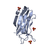
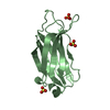
| Title | gamma 2 adaptin EAR domain crystal structure with preS1 site1 peptide NPDWDFN | ||||||
 Components Components |
| ||||||
 Keywords Keywords | PROTEIN TRANSPORT/VIRAL PROTEIN / PROTEIN TRANSPORT-VIRAL PROTEIN COMPLEX / GAE | ||||||
| Function / homology |  Function and homology information Function and homology informationAP-1 adaptor complex / caveolin-mediated endocytosis of virus by host cell / Golgi to vacuole transport / clathrin-cargo adaptor activity / Golgi-associated vesicle / Lysosome Vesicle Biogenesis / transport vesicle / vesicle-mediated transport / intracellular protein transport / endosome membrane ...AP-1 adaptor complex / caveolin-mediated endocytosis of virus by host cell / Golgi to vacuole transport / clathrin-cargo adaptor activity / Golgi-associated vesicle / Lysosome Vesicle Biogenesis / transport vesicle / vesicle-mediated transport / intracellular protein transport / endosome membrane / Golgi membrane / fusion of virus membrane with host endosome membrane / synapse / virion attachment to host cell / virion membrane / Golgi apparatus / membrane Similarity search - Function | ||||||
| Biological species |  Homo sapiens (human) Homo sapiens (human)  HEPATITIS B VIRUS HEPATITIS B VIRUS | ||||||
| Method |  X-RAY DIFFRACTION / SYNCHROTRON / MOLECULAR REPLACEMENT / Resolution: 1.7 Å X-RAY DIFFRACTION / SYNCHROTRON / MOLECULAR REPLACEMENT / Resolution: 1.7 Å | ||||||
 Authors Authors | Juergens, M.C. / Voros, J. / Rautureau, G. / Shepherd, D. / Pye, V.E. / Muldoon, J. / Johnson, C.M. / Ashcroft, A. / Freund, S.M.V. / Ferguson, N. | ||||||
 Citation Citation | Journal: Nat.Chem.Biol. / Year: 2013 Title: The Hepatitis B Virus Pres1 Domain Hijacks Host Trafficking Proteins by Motif Mimicry. Authors: Jurgens, M.C. / Voros, J. / Rautureau, G.J.P. / Shepherd, D.A. / Pye, V.E. / Muldoon, J. / Johnson, C.M. / Ashcroft, A.E. / Freund, S.M.V. / Ferguson, N. | ||||||
| History |
|
- Structure visualization
| Structure viewer | Molecule: MolmilJmol/JSmol |
|---|
- Downloads & links
-Download
| PDBx/mmCIF format | 3zhf.cif.gz | 42.3 KB | Display | PDBx/mmCIF format |
|---|---|---|---|---|
| PDB format | pdb3zhf.ent.gz | 29 KB | Display | PDB format |
| PDBx/mmJSON format | 3zhf.json.gz | Tree view | PDBx/mmJSON format | |
| Others |  Other downloads Other downloads |
-Validation report
| Arichive directory | https://data.pdbj.org/pub/pdb/validation_reports/zh/3zhfftp://data.pdbj.org/pub/pdb/validation_reports/zh/3zhf | HTTPS FTP |
|---|
-Related structure data
| Related structure data |  2ymtC  4bcxSC S: Starting model for refinement C: citing same article ( |
|---|---|
| Similar structure data |


-Links
 PDBj
PDBj


- Assembly
| Deposited unit | 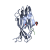
| ||||||||
|---|---|---|---|---|---|---|---|---|---|
| 1 |
| ||||||||
| Unit cell |
|
-Components
| #1: Protein | Mass: 13762.746 Da / Num. of mol.: 1 / Fragment: EAR DOMAIN, RESIDUES 665-785 Source method: isolated from a genetically manipulated source Source: (gene. exp.) Homo sapiens (human) / Production host:  |
|---|---|
| #2: Protein/peptide | Mass: 930.940 Da / Num. of mol.: 1 / Fragment: PRES1 DOMAIN, RESIDUES 85-91 / Source method: obtained synthetically / Details: AMIDATED C-TERMINUS, CARBOXYLATED N-TERMINUS / Source: (synth.) HEPATITIS B VIRUS / References: UniProt: Q67953 |
| #3: Chemical | ChemComp-PEG /   Mass: 106.120 Da / Num. of mol.: 1 / Source method: obtained synthetically / Formula: C4H10O3 Mass: 106.120 Da / Num. of mol.: 1 / Source method: obtained synthetically / Formula: C4H10O3 |
| #4: Chemical | ChemComp-EDO /   Mass: 62.068 Da / Num. of mol.: 1 / Source method: obtained synthetically / Formula: C2H6O2 Mass: 62.068 Da / Num. of mol.: 1 / Source method: obtained synthetically / Formula: C2H6O2 |
| #5: Water | ChemComp-HOH /  Mass: 18.015 Da / Num. of mol.: 84 / Source method: isolated from a natural source / Formula: H2O Mass: 18.015 Da / Num. of mol.: 84 / Source method: isolated from a natural source / Formula: H2O |
| Has protein modification | Y |
| Nonpolymer details | AMINO GROUP (NH2): PEPTIDE C TERMINUS IS AMIDATED |
| Sequence details | GGS IS A CLONING ARTEFACT AT THE PROTEIN N-TERMINUS |
-Experimental details
-Experiment
| Experiment | Method: X-RAY DIFFRACTION / Number of used crystals: 1 |
|---|
- Sample preparation
| Crystal | Density Matthews: 2.51 Å3/Da / Density % sol: 51.07 % / Description: NONE |
|---|---|
| Crystal grow | pH: 6.5 Details: 100 MM MES/IMIDAZOLE PH 6.5, 0.02 M 1,6-HEXANEDIOL, 0.02 M 1-BUTANOL, 0.02 M (RS)-1, 2-PROPANEDIOL, 0.02 M 2-PROPANOL, 0.02 M 1,4-BUTANEDIOL, 0.02 M 1,3-PROPANEDIOL, 10% (W/V) PEG 20000 AND ...Details: 100 MM MES/IMIDAZOLE PH 6.5, 0.02 M 1,6-HEXANEDIOL, 0.02 M 1-BUTANOL, 0.02 M (RS)-1, 2-PROPANEDIOL, 0.02 M 2-PROPANOL, 0.02 M 1,4-BUTANEDIOL, 0.02 M 1,3-PROPANEDIOL, 10% (W/V) PEG 20000 AND 24% (V/V) PEG400 (USING A 2:1 RATIO OF PROTEIN TO MOTHER LIQUOR), PEPTIDE WAS AT ABOUT 50 FOLD MOLAR EXCESS |
-Data collection
| Diffraction | Mean temperature: 93 K |
|---|---|
| Diffraction source | Source: SYNCHROTRON / Site: PETRA III, EMBL c/o DESY  / Beamline: P14 (MX2) / Wavelength: 1.2395 / Beamline: P14 (MX2) / Wavelength: 1.2395 |
| Detector | Type: DECTRIS PILATUS 6M / Detector: PIXEL / Date: Nov 20, 2012 |
| Radiation | Protocol: SINGLE WAVELENGTH / Monochromatic (M) / Laue (L): M / Scattering type: x-ray |
| Radiation wavelength | Wavelength: 1.2395 Å / Relative weight: 1 |
| Reflection | Resolution: 1.7→37.46 Å / Num. obs: 15669 / % possible obs: 98.8 % / Observed criterion σ(I): 2.7 / Redundancy: 4.1 % / Biso Wilson estimate: 20.34 Å2 / Rmerge(I) obs: 0.05 / Net I/σ(I): 17.3 |
| Reflection shell | Resolution: 1.7→1.79 Å / Redundancy: 4.2 % / Rmerge(I) obs: 0.42 / Mean I/σ(I) obs: 2.7 / % possible all: 99.1 |
- Processing
| Software |
| |||||||||||||||||||||||||||||||||||||||||||||||||
|---|---|---|---|---|---|---|---|---|---|---|---|---|---|---|---|---|---|---|---|---|---|---|---|---|---|---|---|---|---|---|---|---|---|---|---|---|---|---|---|---|---|---|---|---|---|---|---|---|---|---|
| Refinement | Method to determine structure: MOLECULAR REPLACEMENT Starting model: PDB ENTRY 4BCX Resolution: 1.7→35.298 Å / SU ML: 0.18 / σ(F): 1.34 / Phase error: 23.82 / Stereochemistry target values: ML
| |||||||||||||||||||||||||||||||||||||||||||||||||
| Solvent computation | Shrinkage radii: 0.9 Å / VDW probe radii: 1.11 Å / Solvent model: FLAT BULK SOLVENT MODEL | |||||||||||||||||||||||||||||||||||||||||||||||||
| Refinement step | Cycle: LAST / Resolution: 1.7→35.298 Å
| |||||||||||||||||||||||||||||||||||||||||||||||||
| Refine LS restraints |
| |||||||||||||||||||||||||||||||||||||||||||||||||
| LS refinement shell |
|
