 Movie
Movie Controller

[English] 日本語
 Yorodumi
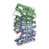
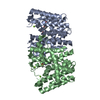
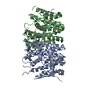
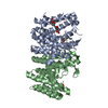
- PDB-3zcd: Native structure of Farnesyl Pyrophosphate Synthase from Pseudomo... -
+ Open data

- Basic information
| Entry | Database: PDB / ID: 3zcd | ||||||
|---|---|---|---|---|---|---|---|
| Title | Native structure of Farnesyl Pyrophosphate Synthase from Pseudomonas aeruginosa PA01. | ||||||
 Components Components | GERANYLTRANSTRANSFERASE | ||||||
 Keywords Keywords | TRANSFERASE / FPPS | ||||||
| Function / homology |  Function and homology information Function and homology informationprenyltransferase activity / phospholipid biosynthetic process / terpenoid biosynthetic process / metal ion binding / cytoplasm Similarity search - Function | ||||||
| Biological species |  PSEUDOMONAS AERUGINOSA PAO1 (bacteria) PSEUDOMONAS AERUGINOSA PAO1 (bacteria) | ||||||
| Method |  X-RAY DIFFRACTION / SYNCHROTRON / MOLECULAR REPLACEMENT / Resolution: 1.55 Å X-RAY DIFFRACTION / SYNCHROTRON / MOLECULAR REPLACEMENT / Resolution: 1.55 Å | ||||||
 Authors Authors | Schmidberger, J.W. / Schnell, R. / Schneider, G. | ||||||
 Citation Citation | Journal: Acta Crystallogr.,Sect.D / Year: 2015 Title: Structural Characterization of Substrate and Inhibitor Binding to Farnesyl Pyrophosphate Synthase from Pseudomonas Aeruginosa. Authors: Schmidberger, J.W. / Schnell, R. / Schneider, G. | ||||||
| History |
|
- Structure visualization
| Structure viewer | Molecule: MolmilJmol/JSmol |
|---|
- Downloads & links
-Download
| PDBx/mmCIF format | 3zcd.cif.gz | 238.2 KB | Display | PDBx/mmCIF format |
|---|---|---|---|---|
| PDB format | pdb3zcd.ent.gz | 193.5 KB | Display | PDB format |
| PDBx/mmJSON format | 3zcd.json.gz | Tree view | PDBx/mmJSON format | |
| Others |  Other downloads Other downloads |
-Validation report
| Arichive directory | https://data.pdbj.org/pub/pdb/validation_reports/zc/3zcdftp://data.pdbj.org/pub/pdb/validation_reports/zc/3zcd | HTTPS FTP |
|---|
-Related structure data
| Related structure data |  3zl6C  3zmbC  3zmcC  3zouC  4umjC  3ljiS S: Starting model for refinement C: citing same article ( |
|---|---|
| Similar structure data |


-Links
 PDBj
PDBj
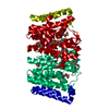
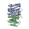
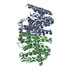
- Assembly
| Deposited unit | 
| |||||||||
|---|---|---|---|---|---|---|---|---|---|---|
| 1 |
| |||||||||
| Unit cell |
| |||||||||
| Components on special symmetry positions |
|
-Components
| #1: Protein | Mass: 31588.836 Da / Num. of mol.: 2 Source method: isolated from a genetically manipulated source Source: (gene. exp.) PSEUDOMONAS AERUGINOSA PAO1 (bacteria) / Production host: References: UniProt: Q9HWY4, (2E,6E)-farnesyl diphosphate synthase #2: Water | ChemComp-HOH / |  Mass: 18.015 Da / Num. of mol.: 559 / Source method: isolated from a natural source / Formula: H2O Mass: 18.015 Da / Num. of mol.: 559 / Source method: isolated from a natural source / Formula: H2OSequence details | THERE IS AN N-TERMINAL ADDITIONAL | |
|---|
-Experimental details
-Experiment
| Experiment | Method: X-RAY DIFFRACTION / Number of used crystals: 1 |
|---|
- Sample preparation
| Crystal | Density Matthews: 2 Å3/Da / Density % sol: 39 % / Description: NONE |
|---|---|
| Crystal grow | pH: 6.5 Details: 25% PEG3350, 0.2 M NAF, 0.1 M BISTRISPROPANE PH 6.5 |
-Data collection
| Diffraction | Mean temperature: 100 K |
|---|---|
| Diffraction source | Source: SYNCHROTRON / Site: ESRF  / Beamline: ID14-1 / Wavelength: 0.9334 / Beamline: ID14-1 / Wavelength: 0.9334 |
| Detector | Type: ADSC QUANTUM 210 / Detector: CCD / Date: Dec 11, 2010 |
| Radiation | Protocol: SINGLE WAVELENGTH / Monochromatic (M) / Laue (L): M / Scattering type: x-ray |
| Radiation wavelength | Wavelength: 0.9334 Å / Relative weight: 1 |
| Reflection | Resolution: 1.55→65.54 Å / Num. obs: 78917 / % possible obs: 99.5 % / Observed criterion σ(I): 2 / Redundancy: 3.7 % / Biso Wilson estimate: 14.6 Å2 / Rmerge(I) obs: 0.1 / Net I/σ(I): 7.2 |
| Reflection shell | Resolution: 1.55→1.63 Å / Redundancy: 3.4 % / Rmerge(I) obs: 0.36 / Mean I/σ(I) obs: 3.4 / % possible all: 99.4 |
- Processing
| Software |
| ||||||||||||||||||||||||||||||||||||||||||||||||||||||||||||||||||||||||||||||||||||||||||||||||||||||||||||||||||||||||||||||||||||||||||||||||||||||||||||||||||||||||||||||||||||||
|---|---|---|---|---|---|---|---|---|---|---|---|---|---|---|---|---|---|---|---|---|---|---|---|---|---|---|---|---|---|---|---|---|---|---|---|---|---|---|---|---|---|---|---|---|---|---|---|---|---|---|---|---|---|---|---|---|---|---|---|---|---|---|---|---|---|---|---|---|---|---|---|---|---|---|---|---|---|---|---|---|---|---|---|---|---|---|---|---|---|---|---|---|---|---|---|---|---|---|---|---|---|---|---|---|---|---|---|---|---|---|---|---|---|---|---|---|---|---|---|---|---|---|---|---|---|---|---|---|---|---|---|---|---|---|---|---|---|---|---|---|---|---|---|---|---|---|---|---|---|---|---|---|---|---|---|---|---|---|---|---|---|---|---|---|---|---|---|---|---|---|---|---|---|---|---|---|---|---|---|---|---|---|---|
| Refinement | Method to determine structure: MOLECULAR REPLACEMENT Starting model: PDB ENTRY 3LJI Resolution: 1.55→25.84 Å / Cor.coef. Fo:Fc: 0.971 / Cor.coef. Fo:Fc free: 0.95 / SU B: 4.601 / SU ML: 0.07 / Cross valid method: THROUGHOUT / ESU R: 0.091 / ESU R Free: 0.081 / Stereochemistry target values: MAXIMUM LIKELIHOOD Details: HYDROGENS HAVE BEEN ADDED IN THE RIDING POSITIONS. U VALUES REFINED INDIVIDUALLY
| ||||||||||||||||||||||||||||||||||||||||||||||||||||||||||||||||||||||||||||||||||||||||||||||||||||||||||||||||||||||||||||||||||||||||||||||||||||||||||||||||||||||||||||||||||||||
| Solvent computation | Ion probe radii: 0.8 Å / Shrinkage radii: 0.8 Å / VDW probe radii: 1.2 Å / Solvent model: MASK | ||||||||||||||||||||||||||||||||||||||||||||||||||||||||||||||||||||||||||||||||||||||||||||||||||||||||||||||||||||||||||||||||||||||||||||||||||||||||||||||||||||||||||||||||||||||
| Displacement parameters | Biso mean: 22.379 Å2
| ||||||||||||||||||||||||||||||||||||||||||||||||||||||||||||||||||||||||||||||||||||||||||||||||||||||||||||||||||||||||||||||||||||||||||||||||||||||||||||||||||||||||||||||||||||||
| Refinement step | Cycle: LAST / Resolution: 1.55→25.84 Å
| ||||||||||||||||||||||||||||||||||||||||||||||||||||||||||||||||||||||||||||||||||||||||||||||||||||||||||||||||||||||||||||||||||||||||||||||||||||||||||||||||||||||||||||||||||||||
| Refine LS restraints |
|