 Movie
Movie Controller

[English] 日本語
 Yorodumi
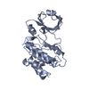
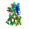
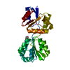
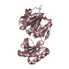
- PDB-3x37: Crystal structure of the N-terminal domain of Sld7 in complex wit... -
+ Open data

- Basic information
| Entry | Database: PDB / ID: 3x37 | ||||||
|---|---|---|---|---|---|---|---|
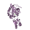
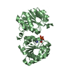
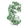
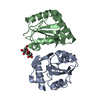
| Title | Crystal structure of the N-terminal domain of Sld7 in complex with Sld3 | ||||||
 Components Components |
| ||||||
 Keywords Keywords | REPLICATION REGULATOR / Beta-barrel | ||||||
| Function / homology |  Function and homology information Function and homology informationDNA replication preinitiation complex / DNA replication initiation / spindle pole / DNA replication / nucleus Similarity search - Function | ||||||
| Biological species |  Zygosaccharomyces rouxii CBS 732 (yeast) Zygosaccharomyces rouxii CBS 732 (yeast) | ||||||
| Method |  X-RAY DIFFRACTION / SYNCHROTRON / SAD / Resolution: 2.35 Å X-RAY DIFFRACTION / SYNCHROTRON / SAD / Resolution: 2.35 Å | ||||||
 Authors Authors | Itou, H. / Araki, H. / Shirakihara, Y. | ||||||
 Citation Citation | Journal: Acta Crystallogr.,Sect.D / Year: 2015 Title: The quaternary structure of the eukaryotic DNA replication proteins Sld7 and Sld3. Authors: Itou, H. / Shirakihara, Y. / Araki, H. | ||||||
| History |
|
- Structure visualization
| Structure viewer | Molecule: MolmilJmol/JSmol |
|---|
- Downloads & links
-Download
| PDBx/mmCIF format | 3x37.cif.gz | 66.1 KB | Display | PDBx/mmCIF format |
|---|---|---|---|---|
| PDB format | pdb3x37.ent.gz | 48.7 KB | Display | PDB format |
| PDBx/mmJSON format | 3x37.json.gz | Tree view | PDBx/mmJSON format | |
| Others |  Other downloads Other downloads |
-Validation report
| Arichive directory | https://data.pdbj.org/pub/pdb/validation_reports/x3/3x37ftp://data.pdbj.org/pub/pdb/validation_reports/x3/3x37 | HTTPS FTP |
|---|
-Related structure data


-Links
 PDBj
PDBj- Assembly
| Deposited unit | 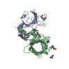
| ||||||||
|---|---|---|---|---|---|---|---|---|---|
| 1 |
| ||||||||
| Unit cell |
|
-Components
| #1: Protein | Mass: 14751.167 Da / Num. of mol.: 1 / Fragment: N-terminal domain (UNP RESIDUES 1-115) Source method: isolated from a genetically manipulated source Source: (gene. exp.) Zygosaccharomyces rouxii CBS 732 (yeast)Strain: ATCC 2623 / CBS 732 / NBRC 1130 / NCYC 568 / NRRL Y-229 Gene: sld3, ZYRO0C14696g / Plasmid: pETduet1 / Production host:  | ||||
|---|---|---|---|---|---|
| #2: Protein | Mass: 15566.771 Da / Num. of mol.: 1 / Fragment: N-terminal domain (UNP RESIDUES 1-133) Source method: isolated from a genetically manipulated source Source: (gene. exp.) Zygosaccharomyces rouxii CBS 732 (yeast)Strain: ATCC 2623 / CBS 732 / NBRC 1130 / NCYC 568 / NRRL Y-229 Gene: SLD7, ZYRO0B16016g / Plasmid: pETduet1 / Production host: | ||||
| #3: Chemical |   Mass: 92.094 Da / Num. of mol.: 2 / Source method: obtained synthetically / Formula: C3H8O3 Mass: 92.094 Da / Num. of mol.: 2 / Source method: obtained synthetically / Formula: C3H8O3#4: Water | ChemComp-HOH / |  Mass: 18.015 Da / Num. of mol.: 81 / Source method: isolated from a natural source / Formula: H2O Mass: 18.015 Da / Num. of mol.: 81 / Source method: isolated from a natural source / Formula: H2OHas protein modification | Y | |
-Experimental details
-Experiment
| Experiment | Method: X-RAY DIFFRACTION / Number of used crystals: 1 |
|---|
- Sample preparation
| Crystal | Density Matthews: 3.09 Å3/Da / Density % sol: 60.23 % |
|---|---|
| Crystal grow | Temperature: 293 K / Method: vapor diffusion, hanging drop / pH: 8.8 Details: 0.1M CHES-NaOH, 0.8M Na-Citrate, pH 8.8, VAPOR DIFFUSION, HANGING DROP, temperature 293K |
-Data collection
| Diffraction | Mean temperature: 100 K |
|---|---|
| Diffraction source | Source: SYNCHROTRON / Site: Photon Factory  / Beamline: BL-17A / Wavelength: 0.97865 Å / Beamline: BL-17A / Wavelength: 0.97865 Å |
| Detector | Type: DECTRIS PILATUS 6M / Detector: PIXEL / Date: Dec 11, 2014 |
| Radiation | Monochromator: Si (111) / Protocol: SINGLE WAVELENGTH / Monochromatic (M) / Laue (L): M / Scattering type: x-ray |
| Radiation wavelength | Wavelength: 0.97865 Å / Relative weight: 1 |
| Reflection | Resolution: 2.35→34.14 Å / Num. all: 15828 / Num. obs: 15781 / % possible obs: 99.7 % / Observed criterion σ(F): 0 / Observed criterion σ(I): 0 / Redundancy: 5 % / Biso Wilson estimate: 41.7 Å2 |
| Reflection shell | Resolution: 2.35→2.48 Å / Redundancy: 4.6 % / Mean I/σ(I) obs: 3.3 / Num. unique all: 2252 / % possible all: 99.7 |
- Processing
| Software |
| |||||||||||||||||||||||||||||||||||
|---|---|---|---|---|---|---|---|---|---|---|---|---|---|---|---|---|---|---|---|---|---|---|---|---|---|---|---|---|---|---|---|---|---|---|---|---|
| Refinement | Method to determine structure: SAD / Resolution: 2.35→19.98 Å / SU ML: 0.29 / σ(F): 1.36 / Phase error: 30.14 / Stereochemistry target values: ML
| |||||||||||||||||||||||||||||||||||
| Solvent computation | Shrinkage radii: 0.9 Å / VDW probe radii: 1.11 Å / Solvent model: FLAT BULK SOLVENT MODEL | |||||||||||||||||||||||||||||||||||
| Refinement step | Cycle: LAST / Resolution: 2.35→19.98 Å
| |||||||||||||||||||||||||||||||||||
| Refine LS restraints |
| |||||||||||||||||||||||||||||||||||
| LS refinement shell | Refine-ID: X-RAY DIFFRACTION / Total num. of bins used: 6 / % reflection obs: 100 %
|
