 Movie
Movie Controller

[English] 日本語
 Yorodumi
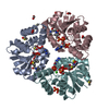
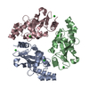
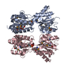
- PDB-3x1k: crystal structure of Phosphoapantetheine adenylyltransferase PPAT... -
+ Open data

- Basic information
| Entry | Database: PDB / ID: 3x1k | ||||||
|---|---|---|---|---|---|---|---|
| Title | crystal structure of Phosphoapantetheine adenylyltransferase PPAT/CoaD with AMP-PNP from Pseudomonas aerugonosa | ||||||
 Components Components | Phosphopantetheine adenylyltransferase | ||||||
 Keywords Keywords | TRANSFERASE / Rossmann Fold | ||||||
| Function / homology |  Function and homology information Function and homology information[citrate (pro-3S)-lyase] ligase activity / pantetheine-phosphate adenylyltransferase / pantetheine-phosphate adenylyltransferase activity / coenzyme A biosynthetic process / ATP binding / cytoplasm Similarity search - Function | ||||||
| Biological species |  Pseudomonas aeruginosa 2192 (bacteria) Pseudomonas aeruginosa 2192 (bacteria) | ||||||
| Method |  X-RAY DIFFRACTION / SYNCHROTRON / MOLECULAR REPLACEMENT / Resolution: 2.547 Å X-RAY DIFFRACTION / SYNCHROTRON / MOLECULAR REPLACEMENT / Resolution: 2.547 Å | ||||||
 Authors Authors | Chatterjee, R. / Datta, S. | ||||||
 Citation Citation | Journal: Biochim.Biophys.Acta / Year: 2016 Title: Transition of phosphopantetheine adenylyltransferase from catalytic to allosteric state is characterized by ternary complex formation in Pseudomonas aeruginosa Authors: Chatterjee, R. / Mondal, A. / Basu, A. / Datta, S. | ||||||
| History |
|
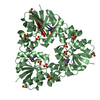
- Structure visualization
| Structure viewer | Molecule: MolmilJmol/JSmol |
|---|
- Downloads & links
-Download
| PDBx/mmCIF format | 3x1k.cif.gz | 201.7 KB | Display | PDBx/mmCIF format |
|---|---|---|---|---|
| PDB format | pdb3x1k.ent.gz | 162.3 KB | Display | PDB format |
| PDBx/mmJSON format | 3x1k.json.gz | Tree view | PDBx/mmJSON format | |
| Others |  Other downloads Other downloads |
-Validation report
| Arichive directory | https://data.pdbj.org/pub/pdb/validation_reports/x1/3x1kftp://data.pdbj.org/pub/pdb/validation_reports/x1/3x1k | HTTPS FTP |
|---|
-Related structure data
| Related structure data | 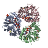 3x1jC 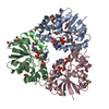 3x1mC  4rukC  1h1tS C: citing same article ( S: Starting model for refinement |
|---|---|
| Similar structure data |


-Links
 PDBj
PDBj


- Assembly
| Deposited unit | 
| ||||||||
|---|---|---|---|---|---|---|---|---|---|
| 1 | 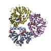
| ||||||||
| 2 | 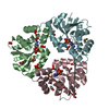
| ||||||||
| Unit cell |
|
-Components
-Protein , 1 types, 6 molecules ABCDEF
| #1: Protein | Mass: 17795.473 Da / Num. of mol.: 6 Source method: isolated from a genetically manipulated source Source: (gene. exp.) Pseudomonas aeruginosa 2192 (bacteria) / Strain: 2192 / Gene: coaD, PA0363, PA2G_04277 / Production host: References: UniProt: A3LHH1, UniProt: A0A0X1KGP2*PLUS, pantetheine-phosphate adenylyltransferase |
|---|
-Non-polymers , 6 types, 149 molecules 


| #2: Chemical | ChemComp-ANP /  Mass: 506.196 Da / Num. of mol.: 6 / Source method: obtained synthetically / Formula: C10H17N6O12P3 / Comment: AMP-PNP, energy-carrying molecule analogue*YM Mass: 506.196 Da / Num. of mol.: 6 / Source method: obtained synthetically / Formula: C10H17N6O12P3 / Comment: AMP-PNP, energy-carrying molecule analogue*YM#3: Chemical | ChemComp-DMS /  Mass: 78.133 Da / Num. of mol.: 16 / Source method: obtained synthetically / Formula: C2H6OS / Comment: DMSO, precipitant*YM Mass: 78.133 Da / Num. of mol.: 16 / Source method: obtained synthetically / Formula: C2H6OS / Comment: DMSO, precipitant*YM#4: Chemical | ChemComp-FMT /  Mass: 46.025 Da / Num. of mol.: 6 / Source method: obtained synthetically / Formula: CH2O2 Mass: 46.025 Da / Num. of mol.: 6 / Source method: obtained synthetically / Formula: CH2O2#5: Chemical |  Mass: 94.971 Da / Num. of mol.: 2 / Source method: obtained synthetically / Formula: PO4 Mass: 94.971 Da / Num. of mol.: 2 / Source method: obtained synthetically / Formula: PO4#6: Chemical |  Mass: 92.094 Da / Num. of mol.: 2 / Source method: obtained synthetically / Formula: C3H8O3 Mass: 92.094 Da / Num. of mol.: 2 / Source method: obtained synthetically / Formula: C3H8O3#7: Water | ChemComp-HOH / | Mass: 18.015 Da / Num. of mol.: 117 / Source method: isolated from a natural source / Formula: H2O |
|---|
-Experimental details
-Experiment
| Experiment | Method: X-RAY DIFFRACTION / Number of used crystals: 1 |
|---|
- Sample preparation
| Crystal | Density Matthews: 2.68 Å3/Da / Density % sol: 54.18 % |
|---|---|
| Crystal grow | Temperature: 295 K / Method: vapor diffusion, sitting drop / pH: 6.9 Details: 20% PEG4000, 0.1M HEPES, 5% Isopropanol, 200mM Sodium acetate, pH 6.9, VAPOR DIFFUSION, SITTING DROP, temperature 295K |
-Data collection
| Diffraction | Mean temperature: 73 K | ||||||||||||||||||
|---|---|---|---|---|---|---|---|---|---|---|---|---|---|---|---|---|---|---|---|
| Diffraction source | Source: SYNCHROTRON / Site: RRCAT INDUS-2  / Beamline: PX-BL21 / Wavelength: 0.9748 Å / Beamline: PX-BL21 / Wavelength: 0.9748 Å | ||||||||||||||||||
| Detector | Type: MARMOSAIC 225 mm CCD / Detector: CCD / Date: Apr 12, 2014 | ||||||||||||||||||
| Radiation | Monochromator: Silicon / Protocol: SINGLE WAVELENGTH / Monochromatic (M) / Laue (L): M / Scattering type: x-ray | ||||||||||||||||||
| Radiation wavelength | Wavelength: 0.9748 Å / Relative weight: 1 | ||||||||||||||||||
| Reflection | Resolution: 2.547→39.745 Å / Num. all: 37216 / Num. obs: 36472 / % possible obs: 98 % / Observed criterion σ(F): 1.8 / Observed criterion σ(I): 1.8 | ||||||||||||||||||
| Reflection shell |
|
- Processing
| Software |
| |||||||||||||||||||||||||||||||||||||||||||||||||||||||||||||||||||||||||||||||||||||||||||||||||||||||||
|---|---|---|---|---|---|---|---|---|---|---|---|---|---|---|---|---|---|---|---|---|---|---|---|---|---|---|---|---|---|---|---|---|---|---|---|---|---|---|---|---|---|---|---|---|---|---|---|---|---|---|---|---|---|---|---|---|---|---|---|---|---|---|---|---|---|---|---|---|---|---|---|---|---|---|---|---|---|---|---|---|---|---|---|---|---|---|---|---|---|---|---|---|---|---|---|---|---|---|---|---|---|---|---|---|---|---|
| Refinement | Method to determine structure: MOLECULAR REPLACEMENT Starting model: 1H1T Resolution: 2.547→39.745 Å / SU ML: 0.35 / σ(F): 1.34 / Phase error: 24.58 / Stereochemistry target values: ML
| |||||||||||||||||||||||||||||||||||||||||||||||||||||||||||||||||||||||||||||||||||||||||||||||||||||||||
| Solvent computation | Shrinkage radii: 0.9 Å / VDW probe radii: 1.11 Å / Solvent model: FLAT BULK SOLVENT MODEL | |||||||||||||||||||||||||||||||||||||||||||||||||||||||||||||||||||||||||||||||||||||||||||||||||||||||||
| Refinement step | Cycle: LAST / Resolution: 2.547→39.745 Å
| |||||||||||||||||||||||||||||||||||||||||||||||||||||||||||||||||||||||||||||||||||||||||||||||||||||||||
| Refine LS restraints |
| |||||||||||||||||||||||||||||||||||||||||||||||||||||||||||||||||||||||||||||||||||||||||||||||||||||||||
| LS refinement shell |
|
