 Movie
Movie Controller


+ Open data

- Basic information
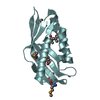
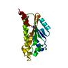
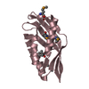
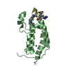
| Entry | Database: PDB / ID: 3wz5 | ||||||
|---|---|---|---|---|---|---|---|
| Title | Structure of the periplasmic domain of DotI (crystal form II) | ||||||
 Components Components | DotI | ||||||
 Keywords Keywords | UNKNOWN FUNCTION / type IVB secretion | ||||||
| Function / homology | Type-IV b secretion system, inner-membrane complex component / Type-IV b secretion system, inner-membrane complex component / DotI Function and homology information Function and homology information | ||||||
| Biological species |   Legionella pneumophila (bacteria) Legionella pneumophila (bacteria) | ||||||
| Method |  X-RAY DIFFRACTION / SYNCHROTRON / MOLECULAR REPLACEMENT / Resolution: 3.5 Å X-RAY DIFFRACTION / SYNCHROTRON / MOLECULAR REPLACEMENT / Resolution: 3.5 Å | ||||||
 Authors Authors | Kuroda, T. / Kubori, T. / Uchida, Y. / Nagai, H. / Imada, K. | ||||||
 Citation Citation | Journal: Sci Rep / Year: 2015 Title: Molecular and structural analysis of Legionella DotI gives insights into an inner membrane complex essential for type IV secretion Authors: Kuroda, T. / Kubori, T. / Thanh Bui, X. / Hyakutake, A. / Uchida, Y. / Imada, K. / Nagai, H. | ||||||
| History |
|
- Structure visualization
| Structure viewer | Molecule: MolmilJmol/JSmol |
|---|
- Downloads & links
-Download
| PDBx/mmCIF format | 3wz5.cif.gz | 113.8 KB | Display | PDBx/mmCIF format |
|---|---|---|---|---|
| PDB format | pdb3wz5.ent.gz | 90.8 KB | Display | PDB format |
| PDBx/mmJSON format | 3wz5.json.gz | Tree view | PDBx/mmJSON format | |
| Others |  Other downloads Other downloads |
-Validation report
| Arichive directory | https://data.pdbj.org/pub/pdb/validation_reports/wz/3wz5ftp://data.pdbj.org/pub/pdb/validation_reports/wz/3wz5 | HTTPS FTP |
|---|
-Related structure data
| Related structure data |  3wz3C 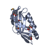 3wz4SC C: citing same article ( S: Starting model for refinement |
|---|---|
| Similar structure data |


-Links
 PDBj
PDBj- Assembly
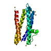
| Deposited unit | 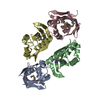
| ||||||||
|---|---|---|---|---|---|---|---|---|---|
| 1 | 
| ||||||||
| 2 | 
| ||||||||
| 3 | 
| ||||||||
| 4 | 
| ||||||||
| Unit cell |
| ||||||||
| Details | AUTHOR DETERMINED BIOLOGICAL UNIT: UNKNOWN |
-Components
| #1: Protein | Mass: 15725.213 Da / Num. of mol.: 4 / Fragment: periplasmic domain, UNP residues 73-212 Source method: isolated from a genetically manipulated source Source: (gene. exp.) Legionella pneumophila (bacteria) / Gene: dotI, icmL / Plasmid: pET15b / Production host: Has protein modification | Y | |
|---|
-Experimental details
-Experiment
| Experiment | Method: X-RAY DIFFRACTION / Number of used crystals: 1 |
|---|
- Sample preparation
| Crystal | Density Matthews: 4.05 Å3/Da / Density % sol: 69.61 % |
|---|---|
| Crystal grow | Temperature: 277 K / Method: vapor diffusion, sitting drop / pH: 7.5 Details: 0.1M HEPES pH7.5, 0.2M NaCl, 40% PEG 300, VAPOR DIFFUSION, SITTING DROP, temperature 277K |
-Data collection
| Diffraction | Mean temperature: 35 K |
|---|---|
| Diffraction source | Source: SYNCHROTRON / Site: SPring-8  / Beamline: BL41XU / Wavelength: 0.97923 Å / Beamline: BL41XU / Wavelength: 0.97923 Å |
| Detector | Type: RAYONIX MX-225 / Detector: CCD / Date: Nov 1, 2010 |
| Radiation | Monochromator: Double-crystal monochromator / Protocol: SINGLE WAVELENGTH / Monochromatic (M) / Laue (L): M / Scattering type: x-ray |
| Radiation wavelength | Wavelength: 0.97923 Å / Relative weight: 1 |
| Reflection | Resolution: 3.5→81.4 Å / Num. all: 13479 / Num. obs: 13479 / % possible obs: 100 % / Observed criterion σ(F): 0 / Observed criterion σ(I): 0 / Redundancy: 11.5 % / Biso Wilson estimate: 11.9 Å2 / Rmerge(I) obs: 0.125 / Rsym value: 0.125 / Net I/σ(I): 16.7 |
| Reflection shell | Resolution: 3.5→3.69 Å / Redundancy: 11.8 % / Rmerge(I) obs: 0.242 / Mean I/σ(I) obs: 10.4 / Num. unique all: 1913 / % possible all: 100 |
- Processing
| Software |
| ||||||||||||||||||||||||||||||||||||
|---|---|---|---|---|---|---|---|---|---|---|---|---|---|---|---|---|---|---|---|---|---|---|---|---|---|---|---|---|---|---|---|---|---|---|---|---|---|
| Refinement | Method to determine structure: MOLECULAR REPLACEMENT Starting model: 3WZ4 Resolution: 3.5→72.836 Å / SU ML: 0.63 / σ(F): 1.5 / Phase error: 18.89 / Stereochemistry target values: ML
| ||||||||||||||||||||||||||||||||||||
| Solvent computation | Shrinkage radii: 0.83 Å / VDW probe radii: 1.1 Å / Solvent model: FLAT BULK SOLVENT MODEL / Bsol: 8.86 Å2 / ksol: 0.327 e/Å3 | ||||||||||||||||||||||||||||||||||||
| Displacement parameters |
| ||||||||||||||||||||||||||||||||||||
| Refinement step | Cycle: LAST / Resolution: 3.5→72.836 Å
| ||||||||||||||||||||||||||||||||||||
| Refine LS restraints |
| ||||||||||||||||||||||||||||||||||||
| LS refinement shell | Refine-ID: X-RAY DIFFRACTION / Total num. of bins used: 5
|
