 Movie
Movie Controller

[English] 日本語
 Yorodumi
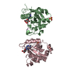
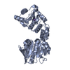
- PDB-3wtz: Crystal structure of ETS-1 DNA binding and autoinhibitory domains... -
+ Open data

- Basic information
| Entry | Database: PDB / ID: 3wtz | ||||||
|---|---|---|---|---|---|---|---|
| Title | Crystal structure of ETS-1 DNA binding and autoinhibitory domains (276-441) | ||||||
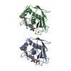
 Components Components | Protein C-ets-1 | ||||||
 Keywords Keywords | TRANSCRIPTION / ETS-1 / AUTOINHIBITION / ETS DOMAIN / DNA-BINDING / ISOPEPTIDE BOND / NUCLEUS / PHOSPHOPROTEIN / PROTO-ONCOGENE / TRANSCRIPTION REGULATION | ||||||
| Function / homology |  Function and homology information Function and homology informationPML body organization / positive regulation of leukocyte adhesion to vascular endothelial cell / negative regulation of cell cycle / regulation of angiogenesis / positive regulation of blood vessel endothelial cell migration / positive regulation of endothelial cell migration / positive regulation of erythrocyte differentiation / transcription corepressor binding / cell motility / positive regulation of miRNA transcription ...PML body organization / positive regulation of leukocyte adhesion to vascular endothelial cell / negative regulation of cell cycle / regulation of angiogenesis / positive regulation of blood vessel endothelial cell migration / positive regulation of endothelial cell migration / positive regulation of erythrocyte differentiation / transcription corepressor binding / cell motility / positive regulation of miRNA transcription / Oncogene Induced Senescence / positive regulation of angiogenesis / positive regulation of inflammatory response / transcription by RNA polymerase II / DNA-binding transcription activator activity, RNA polymerase II-specific / regulation of apoptotic process / nucleic acid binding / RNA polymerase II-specific DNA-binding transcription factor binding / DNA-binding transcription factor activity, RNA polymerase II-specific / cell differentiation / transcription cis-regulatory region binding / immune response / RNA polymerase II cis-regulatory region sequence-specific DNA binding / DNA-binding transcription factor activity / negative regulation of cell population proliferation / response to antibiotic / positive regulation of gene expression / regulation of transcription by RNA polymerase II / positive regulation of DNA-templated transcription / chromatin / DNA-templated transcription / positive regulation of transcription by RNA polymerase II / DNA binding / nucleoplasm / identical protein binding / nucleus / cytoplasm Similarity search - Function | ||||||
| Biological species |  Homo sapiens (human) Homo sapiens (human) | ||||||
| Method |  X-RAY DIFFRACTION / SYNCHROTRON / MOLECULAR REPLACEMENT / Resolution: 2.61 Å X-RAY DIFFRACTION / SYNCHROTRON / MOLECULAR REPLACEMENT / Resolution: 2.61 Å | ||||||
 Authors Authors | Shiina, M. / Hamada, K. / Ogata, K. | ||||||
 Citation Citation | Journal: J.Mol.Biol. / Year: 2015 Title: A novel allosteric mechanism on protein-DNA interactions underlying the phosphorylation-dependent regulation of Ets1 target gene expressions. Authors: Shiina, M. / Hamada, K. / Inoue-Bungo, T. / Shimamura, M. / Uchiyama, A. / Baba, S. / Sato, K. / Yamamoto, M. / Ogata, K. | ||||||
| History |
|
- Structure visualization
| Structure viewer | Molecule: MolmilJmol/JSmol |
|---|
- Downloads & links
-Download
| PDBx/mmCIF format | 3wtz.cif.gz | 68.1 KB | Display | PDBx/mmCIF format |
|---|---|---|---|---|
| PDB format | pdb3wtz.ent.gz | 50.3 KB | Display | PDB format |
| PDBx/mmJSON format | 3wtz.json.gz | Tree view | PDBx/mmJSON format | |
| Others |  Other downloads Other downloads |
-Validation report
| Arichive directory | https://data.pdbj.org/pub/pdb/validation_reports/wt/3wtzftp://data.pdbj.org/pub/pdb/validation_reports/wt/3wtz | HTTPS FTP |
|---|
-Related structure data
| Related structure data |  3wtsC  3wttC  3wtuC  3wtvC 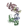 3wtwC 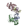 3wtxC  3wtyC  3wu0C  3wu1C 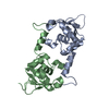 1gvjS C: citing same article ( S: Starting model for refinement |
|---|---|
| Similar structure data |


-Links
 PDBj
PDBj- Assembly
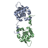
| Deposited unit | 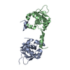
| ||||||||
|---|---|---|---|---|---|---|---|---|---|
| 1 |
| ||||||||
| Unit cell |
|
-Components
| #1: Protein | Mass: 19216.732 Da / Num. of mol.: 2 / Fragment: UNP RESIDUES 276-441 Source method: isolated from a genetically manipulated source Source: (gene. exp.) Homo sapiens (human) / Gene: ETS1, EWSR2 / Plasmid: PET23A / Production host:  #2: Water | ChemComp-HOH / |  Mass: 18.015 Da / Num. of mol.: 22 / Source method: isolated from a natural source / Formula: H2O Mass: 18.015 Da / Num. of mol.: 22 / Source method: isolated from a natural source / Formula: H2O |
|---|
-Experimental details
-Experiment
| Experiment | Method: X-RAY DIFFRACTION / Number of used crystals: 1 |
|---|
- Sample preparation
| Crystal | Density Matthews: 2.65 Å3/Da / Density % sol: 53.62 % |
|---|---|
| Crystal grow | Method: vapor diffusion, sitting drop / pH: 7.8 Details: 9% MPD, 6% PEG 6000, 0.1M HEPES, pH 7.8, VAPOR DIFFUSION, SITTING DROP |
-Data collection
| Diffraction | Mean temperature: 100 K |
|---|---|
| Diffraction source | Source: SYNCHROTRON / Site: Photon Factory  / Beamline: AR-NW12A / Wavelength: 1 Å / Beamline: AR-NW12A / Wavelength: 1 Å |
| Detector | Type: ADSC QUANTUM 210 / Detector: CCD / Date: Mar 10, 2006 |
| Radiation | Protocol: SINGLE WAVELENGTH / Monochromatic (M) / Laue (L): M / Scattering type: x-ray |
| Radiation wavelength | Wavelength: 1 Å / Relative weight: 1 |
| Reflection | Resolution: 2.6→50 Å / Num. obs: 11974 / % possible obs: 99.2 % / Redundancy: 5.3 % / Rmerge(I) obs: 0.059 / Net I/σ(I): 15.7 |
| Reflection shell | Resolution: 2.6→2.69 Å / Redundancy: 3.5 % / Rmerge(I) obs: 0.325 / % possible all: 96 |
- Processing
| Software |
| ||||||||||||||||||||||||||||||||||||||||||||||||||||||||||||||||||||||||||||||||
|---|---|---|---|---|---|---|---|---|---|---|---|---|---|---|---|---|---|---|---|---|---|---|---|---|---|---|---|---|---|---|---|---|---|---|---|---|---|---|---|---|---|---|---|---|---|---|---|---|---|---|---|---|---|---|---|---|---|---|---|---|---|---|---|---|---|---|---|---|---|---|---|---|---|---|---|---|---|---|---|---|---|
| Refinement | Method to determine structure: MOLECULAR REPLACEMENT Starting model: PDB ENTRY 1GVJ Resolution: 2.61→36.42 Å / Rfactor Rfree error: 0.007 / Data cutoff high absF: 821361 / Data cutoff low absF: 0 / Isotropic thermal model: RESTRAINED / Cross valid method: THROUGHOUT / σ(F): 0 / Details: BULK SOLVENT MODEL USED
| ||||||||||||||||||||||||||||||||||||||||||||||||||||||||||||||||||||||||||||||||
| Solvent computation | Solvent model: FLAT MODEL / Bsol: 80.99 Å2 / ksol: 0.4 e/Å3 | ||||||||||||||||||||||||||||||||||||||||||||||||||||||||||||||||||||||||||||||||
| Displacement parameters | Biso mean: 85.2 Å2
| ||||||||||||||||||||||||||||||||||||||||||||||||||||||||||||||||||||||||||||||||
| Refine analyze |
| ||||||||||||||||||||||||||||||||||||||||||||||||||||||||||||||||||||||||||||||||
| Refinement step | Cycle: LAST / Resolution: 2.61→36.42 Å
| ||||||||||||||||||||||||||||||||||||||||||||||||||||||||||||||||||||||||||||||||
| Refine LS restraints |
| ||||||||||||||||||||||||||||||||||||||||||||||||||||||||||||||||||||||||||||||||
| LS refinement shell | Resolution: 2.6→2.76 Å / Rfactor Rfree error: 0.024 / Total num. of bins used: 6
| ||||||||||||||||||||||||||||||||||||||||||||||||||||||||||||||||||||||||||||||||
| Xplor file |
|
