 Movie
Movie Controller

[English] 日本語
 Yorodumi
- PDB-3wrs: Minute virus of mice non-structural protein-1N-terminal nuclease ... -
+ Open data

- Basic information
| Entry | Database: PDB / ID: 3wrs | ||||||
|---|---|---|---|---|---|---|---|
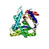
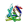
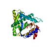
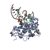
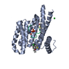
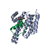
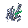
| Title | Minute virus of mice non-structural protein-1N-terminal nuclease domain reveals a unique Zn2+ coordination in the active site pocket and shows a novel mode of DNA recognition at the origin of replication | ||||||
 Components Components | Non-capsid protein NS-1 | ||||||
 Keywords Keywords | REPLICATION / Nuclease activity / Single and double stranded DNA binding and nicking protein | ||||||
| Function / homology |  Function and homology information Function and homology informationrolling hairpin viral DNA replication / symbiont-mediated perturbation of host transcription / symbiont-mediated perturbation of host apoptosis / symbiont-mediated arrest of host cell cycle during G2/M transition / Hydrolases; Acting on ester bonds; Endodeoxyribonucleases producing 5'-phosphomonoesters / symbiont-mediated perturbation of host cell cycle G1/S transition checkpoint / DNA helicase activity / helicase activity / endonuclease activity / DNA helicase ...rolling hairpin viral DNA replication / symbiont-mediated perturbation of host transcription / symbiont-mediated perturbation of host apoptosis / symbiont-mediated arrest of host cell cycle during G2/M transition / Hydrolases; Acting on ester bonds; Endodeoxyribonucleases producing 5'-phosphomonoesters / symbiont-mediated perturbation of host cell cycle G1/S transition checkpoint / DNA helicase activity / helicase activity / endonuclease activity / DNA helicase / DNA replication / host cell nucleus / ATP hydrolysis activity / DNA-templated transcription / DNA binding / ATP binding / metal ion binding Similarity search - Function | ||||||
| Biological species |  Murine minute virus Murine minute virus | ||||||
| Method |  X-RAY DIFFRACTION / SYNCHROTRON / MIR / Resolution: 1.58 Å X-RAY DIFFRACTION / SYNCHROTRON / MIR / Resolution: 1.58 Å | ||||||
 Authors Authors | Tewary, S.K. / Zhao, H. / Tang, L. | ||||||
 Citation Citation | Journal: Virology / Year: 2014 Title: Structures of minute virus of mice replication initiator protein N-terminal domain: Insights into DNA nicking and origin binding. Authors: Tewary, S.K. / Liang, L. / Lin, Z. / Lynn, A. / Cotmore, S.F. / Tattersall, P. / Zhao, H. / Tang, L. | ||||||
| History |
|
- Structure visualization
| Structure viewer | Molecule: MolmilJmol/JSmol |
|---|
- Downloads & links
-Download
| PDBx/mmCIF format | 3wrs.cif.gz | 121.2 KB | Display | PDBx/mmCIF format |
|---|---|---|---|---|
| PDB format | pdb3wrs.ent.gz | 93.5 KB | Display | PDB format |
| PDBx/mmJSON format | 3wrs.json.gz | Tree view | PDBx/mmJSON format | |
| Others |  Other downloads Other downloads |
-Validation report
| Arichive directory | https://data.pdbj.org/pub/pdb/validation_reports/wr/3wrsftp://data.pdbj.org/pub/pdb/validation_reports/wr/3wrs | HTTPS FTP |
|---|
-Related structure data
| Related structure data | 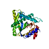 3wrnC  3wroC  3wrqC 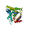 3wrrC  4pp4C 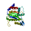 4r94C C: citing same article ( |
|---|---|
| Similar structure data |


-Links
 PDBj
PDBj


- Assembly
| Deposited unit | 
| ||||||||
|---|---|---|---|---|---|---|---|---|---|
| 1 |
| ||||||||
| Unit cell |
|
-Components
| #1: Protein | Mass: 32343.402 Da / Num. of mol.: 1 / Fragment: UNP RESIDUES 1-255 Source method: isolated from a genetically manipulated source Source: (gene. exp.) Murine minute virus / Strain: MVM prototype / Gene: NS1 / Plasmid: pET28a / Production host:  |
|---|---|
| #2: Chemical | ChemComp-CO /   Mass: 58.933 Da / Num. of mol.: 1 / Source method: obtained synthetically / Formula: Co Mass: 58.933 Da / Num. of mol.: 1 / Source method: obtained synthetically / Formula: Co |
| #3: Water | ChemComp-HOH /  Mass: 18.015 Da / Num. of mol.: 169 / Source method: isolated from a natural source / Formula: H2O Mass: 18.015 Da / Num. of mol.: 169 / Source method: isolated from a natural source / Formula: H2O |
-Experimental details
-Experiment
| Experiment | Method: X-RAY DIFFRACTION / Number of used crystals: 1 |
|---|
- Sample preparation
| Crystal | Density Matthews: 2.22 Å3/Da / Density % sol: 44.49 % |
|---|---|
| Crystal grow | Temperature: 293 K / Method: vapor diffusion, hanging drop / pH: 5.4 Details: 2.8M sodium formate, 100mM sodium acetate trihydrate, pH 5.4, VAPOR DIFFUSION, HANGING DROP, temperature 293K |
-Data collection
| Diffraction source | Source: SYNCHROTRON / Site: SSRL  / Beamline: BL12-2 / Wavelength: 0.9795 Å / Beamline: BL12-2 / Wavelength: 0.9795 Å |
|---|---|
| Detector | Type: PSI PILATUS 6M / Detector: PIXEL / Date: Feb 20, 2014 / Details: Mirrors-Rh coated |
| Radiation | Monochromator: Liquid nitrogen-cooled double crystal / Protocol: SINGLE WAVELENGTH / Monochromatic (M) / Laue (L): M / Scattering type: x-ray |
| Radiation wavelength | Wavelength: 0.9795 Å / Relative weight: 1 |
| Reflection | Highest resolution: 1.58 Å / Num. obs: 38421 |
- Processing
| Software |
| ||||||||||||||||||||||||||||||||||||||||||||||||||||||||||||||||||||||||||||||||||||||||||
|---|---|---|---|---|---|---|---|---|---|---|---|---|---|---|---|---|---|---|---|---|---|---|---|---|---|---|---|---|---|---|---|---|---|---|---|---|---|---|---|---|---|---|---|---|---|---|---|---|---|---|---|---|---|---|---|---|---|---|---|---|---|---|---|---|---|---|---|---|---|---|---|---|---|---|---|---|---|---|---|---|---|---|---|---|---|---|---|---|---|---|---|
| Refinement | Method to determine structure: MIR / Resolution: 1.58→34.218 Å / SU ML: 0.16 / σ(F): 1.34 / Phase error: 23.31 / Stereochemistry target values: ML
| ||||||||||||||||||||||||||||||||||||||||||||||||||||||||||||||||||||||||||||||||||||||||||
| Solvent computation | Shrinkage radii: 0.9 Å / VDW probe radii: 1.11 Å / Solvent model: FLAT BULK SOLVENT MODEL | ||||||||||||||||||||||||||||||||||||||||||||||||||||||||||||||||||||||||||||||||||||||||||
| Refinement step | Cycle: LAST / Resolution: 1.58→34.218 Å
| ||||||||||||||||||||||||||||||||||||||||||||||||||||||||||||||||||||||||||||||||||||||||||
| Refine LS restraints |
| ||||||||||||||||||||||||||||||||||||||||||||||||||||||||||||||||||||||||||||||||||||||||||
| LS refinement shell | Refine-ID: X-RAY DIFFRACTION / Total num. of bins used: 14
| ||||||||||||||||||||||||||||||||||||||||||||||||||||||||||||||||||||||||||||||||||||||||||
| Refinement TLS params. | Method: refined / Origin x: 5.0677 Å / Origin y: 42.645 Å / Origin z: 20.7354 Å
| ||||||||||||||||||||||||||||||||||||||||||||||||||||||||||||||||||||||||||||||||||||||||||
| Refinement TLS group | Selection details: CHAIN A |
