 Movie
Movie Controller

[English] 日本語
 Yorodumi
- PDB-3woa: Crystal structure of lambda repressor (1-45) fused with maltose-b... -
+ Open data

- Basic information
| Entry | Database: PDB / ID: 3woa | |||||||||
|---|---|---|---|---|---|---|---|---|---|---|
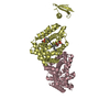
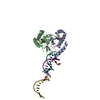
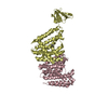
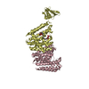
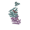
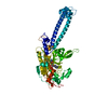
| Title | Crystal structure of lambda repressor (1-45) fused with maltose-binding protein | |||||||||
 Components Components | Repressor protein CI, Maltose-binding periplasmic protein | |||||||||
 Keywords Keywords | DNA BINDING PROTEIN / SUGAR BINDING PROTEIN / LAMBDA REPRESSOR / MALTOSE-BINDING PROTEIN | |||||||||
| Function / homology |  Function and homology information Function and homology informationmaintenance of viral latency / latency-replication decision / positive regulation of viral transcription / : / detection of maltose stimulus / maltose transport complex / carbohydrate transport / carbohydrate transmembrane transporter activity / maltose binding / maltose transport ...maintenance of viral latency / latency-replication decision / positive regulation of viral transcription / : / detection of maltose stimulus / maltose transport complex / carbohydrate transport / carbohydrate transmembrane transporter activity / maltose binding / maltose transport / maltodextrin transmembrane transport / core promoter sequence-specific DNA binding / ATP-binding cassette (ABC) transporter complex, substrate-binding subunit-containing / ATP-binding cassette (ABC) transporter complex / cell chemotaxis / outer membrane-bounded periplasmic space / periplasmic space / DNA damage response / membrane / identical protein binding Similarity search - Function | |||||||||
| Biological species |  Enterobacteria phage lambda (virus) Enterobacteria phage lambda (virus) | |||||||||
| Method |  X-RAY DIFFRACTION / SYNCHROTRON / MOLECULAR REPLACEMENT / Resolution: 2 Å X-RAY DIFFRACTION / SYNCHROTRON / MOLECULAR REPLACEMENT / Resolution: 2 Å | |||||||||
 Authors Authors | Hanazono, Y. / Takeda, K. / Miki, K. | |||||||||
 Citation Citation | Journal: FEBS Open Bio / Year: 2018 Title: Co-translational folding of alpha-helical proteins: structural studies of intermediate-length variants of the lambda repressor. Authors: Hanazono, Y. / Takeda, K. / Miki, K. | |||||||||
| History |
|
- Structure visualization
| Structure viewer | Molecule: MolmilJmol/JSmol |
|---|
- Downloads & links
-Download
| PDBx/mmCIF format | 3woa.cif.gz | 107.1 KB | Display | PDBx/mmCIF format |
|---|---|---|---|---|
| PDB format | pdb3woa.ent.gz | 78.7 KB | Display | PDB format |
| PDBx/mmJSON format | 3woa.json.gz | Tree view | PDBx/mmJSON format | |
| Others |  Other downloads Other downloads |
-Validation report
| Arichive directory | https://data.pdbj.org/pub/pdb/validation_reports/wo/3woaftp://data.pdbj.org/pub/pdb/validation_reports/wo/3woa | HTTPS FTP |
|---|
-Related structure data
| Related structure data |  1anfS S: Starting model for refinement |
|---|---|
| Similar structure data |


-Links
 PDBj
PDBj


- Assembly
| Deposited unit | 
| |||||||||||||||
|---|---|---|---|---|---|---|---|---|---|---|---|---|---|---|---|---|
| 1 |
| |||||||||||||||
| Unit cell |
| |||||||||||||||
| Components on special symmetry positions |
|
-Components
| #1: Protein | Mass: 45916.035 Da / Num. of mol.: 1 / Mutation: N416R Source method: isolated from a genetically manipulated source Details: N-TERMINAL RESIDUES 0-45 FROM REPRESSOR PROTEIN CI (P03034) FUSED WITH MALTOSE-BINDING PERIPLASMIC PROTEIN (P0AEX9) Source: (gene. exp.) Enterobacteria phage lambda (virus), (gene. exp.) Strain: K12 / Gene: malE, b4034, JW3994 / Production host: |
|---|---|
| #2: Polysaccharide | alpha-D-glucopyranose-(1-4)-alpha-D-glucopyranose / alpha-maltose  Source method: isolated from a genetically manipulated source Details: oligosaccharide / References: alpha-maltose |
| #3: Water | ChemComp-HOH /  Mass: 18.015 Da / Num. of mol.: 499 / Source method: isolated from a natural source / Formula: H2O Mass: 18.015 Da / Num. of mol.: 499 / Source method: isolated from a natural source / Formula: H2O |
-Experimental details
-Experiment
| Experiment | Method: X-RAY DIFFRACTION / Number of used crystals: 1 |
|---|
- Sample preparation
| Crystal | Density Matthews: 2.52 Å3/Da / Density % sol: 51.17 % |
|---|---|
| Crystal grow | Temperature: 293 K / Method: vapor diffusion, sitting drop / pH: 7 Details: 1.6M DL-Malic acid, pH 7.0, VAPOR DIFFUSION, SITTING DROP, temperature 293K |
-Data collection
| Diffraction | Mean temperature: 100 K |
|---|---|
| Diffraction source | Source: SYNCHROTRON / Site: SPring-8  / Beamline: BL41XU / Wavelength: 1 Å / Beamline: BL41XU / Wavelength: 1 Å |
| Detector | Type: RAYONIX MX-225 / Detector: CCD / Date: Jun 27, 2012 |
| Radiation | Protocol: SINGLE WAVELENGTH / Monochromatic (M) / Laue (L): M / Scattering type: x-ray |
| Radiation wavelength | Wavelength: 1 Å / Relative weight: 1 |
| Reflection | Resolution: 2→50 Å / Num. obs: 32189 / % possible obs: 99.9 % / Redundancy: 6.8 % / Rsym value: 0.126 / Net I/σ(I): 15.1 |
| Reflection shell | Resolution: 2→2.03 Å / Redundancy: 6.2 % / Mean I/σ(I) obs: 11.3 / Rsym value: 0.179 / % possible all: 99.6 |
- Processing
| Software |
| ||||||||||||||||||||||||||||||||||||||||||||||||||||||||||||||||||||||||||||||
|---|---|---|---|---|---|---|---|---|---|---|---|---|---|---|---|---|---|---|---|---|---|---|---|---|---|---|---|---|---|---|---|---|---|---|---|---|---|---|---|---|---|---|---|---|---|---|---|---|---|---|---|---|---|---|---|---|---|---|---|---|---|---|---|---|---|---|---|---|---|---|---|---|---|---|---|---|---|---|---|
| Refinement | Method to determine structure: MOLECULAR REPLACEMENT Starting model: 1ANF Resolution: 2→34.682 Å / FOM work R set: 0.8805 / SU ML: 0.15 / σ(F): 1.35 / Phase error: 18.12 / Stereochemistry target values: ML
| ||||||||||||||||||||||||||||||||||||||||||||||||||||||||||||||||||||||||||||||
| Solvent computation | Shrinkage radii: 0.9 Å / VDW probe radii: 1.11 Å / Solvent model: FLAT BULK SOLVENT MODEL | ||||||||||||||||||||||||||||||||||||||||||||||||||||||||||||||||||||||||||||||
| Displacement parameters | Biso max: 89.7 Å2 / Biso mean: 18.17 Å2 / Biso min: 4.46 Å2 | ||||||||||||||||||||||||||||||||||||||||||||||||||||||||||||||||||||||||||||||
| Refinement step | Cycle: LAST / Resolution: 2→34.682 Å
| ||||||||||||||||||||||||||||||||||||||||||||||||||||||||||||||||||||||||||||||
| Refine LS restraints |
| ||||||||||||||||||||||||||||||||||||||||||||||||||||||||||||||||||||||||||||||
| LS refinement shell | Refine-ID: X-RAY DIFFRACTION / Total num. of bins used: 12 / % reflection obs: 100 %
|
