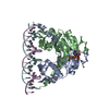
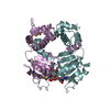
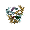
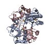
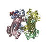
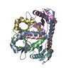
Entry Database : PDB / ID : 3wggTitle Crystal structure of RSP in complex with alpha-NAD+ Redox-sensing transcriptional repressor rex Keywords / / / Function / homology Function Domain/homology Component
/ / / / / / / / / / / / / / / / / / / / / / / / / / / / / / / / Biological species Thermoanaerobacter ethanolicus (bacteria)Method / / / Resolution : 2.1 Å Authors Zheng, Y. / Ko, T.-P. / Guo, R.-T. Journal : Biochem.Biophys.Res.Commun. / Year : 2015Title : Binding mode of the oxidized alpha-anomer of NAD(+) to RSP, a Rex-family repressorAuthors : Zheng, Y. / Ko, T.-P. / Yang, Y. / Shao, W. / Guo, R.-T. History Deposition Aug 5, 2013 Deposition site / Processing site Revision 1.0 Aug 13, 2014 Provider / Type Revision 1.1 Jan 28, 2015 Group Revision 1.2 Nov 8, 2023 Group Data collection / Database references ... Data collection / Database references / Derived calculations / Refinement description Category chem_comp_atom / chem_comp_bond ... chem_comp_atom / chem_comp_bond / database_2 / pdbx_initial_refinement_model / struct_ncs_dom_lim / struct_site Item _database_2.pdbx_DOI / _database_2.pdbx_database_accession ... _database_2.pdbx_DOI / _database_2.pdbx_database_accession / _struct_ncs_dom_lim.beg_auth_comp_id / _struct_ncs_dom_lim.beg_label_asym_id / _struct_ncs_dom_lim.beg_label_comp_id / _struct_ncs_dom_lim.beg_label_seq_id / _struct_ncs_dom_lim.end_auth_comp_id / _struct_ncs_dom_lim.end_label_asym_id / _struct_ncs_dom_lim.end_label_comp_id / _struct_ncs_dom_lim.end_label_seq_id / _struct_site.pdbx_auth_asym_id / _struct_site.pdbx_auth_comp_id / _struct_site.pdbx_auth_seq_id
Show all Show less
 Movie
Movie Controller


 Open data
 Basic information
 Components
 Keywords
 Function and homology information
 Thermoanaerobacter ethanolicus (bacteria)
 X-RAY DIFFRACTION /
  Authors
 Citation
 Structure visualization
 Downloads & links
 Other downloads

 PDBj
PDBj
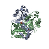
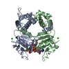
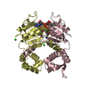
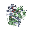
 Assembly
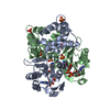
 Mass: 663.425 Da / Num. of mol.: 2 / Source method: obtained synthetically / Formula: C21H27N7O14P2
 Mass: 96.063 Da / Num. of mol.: 6 / Source method: obtained synthetically / Formula: SO4
 Mass: 18.015 Da / Num. of mol.: 418 / Source method: isolated from a natural source / Formula: H2O
 Sample preparation
 / Beamline: BL13B1 / Wavelength: 1 Å
 Processing