 Movie
Movie Controller

[English] 日本語
 Yorodumi
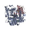
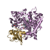
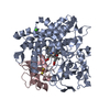
- PDB-3w9c: Crystal structure of the electron transfer complex of cytochrome ... -
+ Open data

- Basic information
| Entry | Database: PDB / ID: 3w9c | ||||||
|---|---|---|---|---|---|---|---|
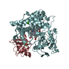
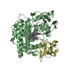
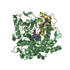
| Title | Crystal structure of the electron transfer complex of cytochrome p450cam with putidaredoxin | ||||||
 Components Components |
| ||||||
 Keywords Keywords | OXIDOREDUCTASE/ELECTRON TRANSPORT / inter-protein electron transfer / OXIDOREDUCTASE-ELECTRON TRANSPORT complex | ||||||
| Function / homology |  Function and homology information Function and homology informationP450-containing electron transport chain / camphor 5-monooxygenase / camphor 5-monooxygenase activity / (+)-camphor catabolic process / 2 iron, 2 sulfur cluster binding / electron transfer activity / iron ion binding / heme binding / metal ion binding / cytosol / cytoplasm Similarity search - Function | ||||||
| Biological species |  Pseudomonas putida (bacteria) Pseudomonas putida (bacteria) | ||||||
| Method |  X-RAY DIFFRACTION / SYNCHROTRON / MOLECULAR REPLACEMENT / Resolution: 2.5 Å X-RAY DIFFRACTION / SYNCHROTRON / MOLECULAR REPLACEMENT / Resolution: 2.5 Å | ||||||
 Authors Authors | Kikui, Y. / Hiruma, Y. / Hass, M.A. / Koteishi, H. / Ubbink, M. / Nojiri, M. | ||||||
 Citation Citation | Journal: J.Mol.Biol. / Year: 2013 Title: The structure of the cytochrome p450cam-putidaredoxin complex determined by paramagnetic NMR spectroscopy and crystallography. Authors: Hiruma, Y. / Hass, M.A. / Kikui, Y. / Liu, W.M. / Olmez, B. / Skinner, S.P. / Blok, A. / Kloosterman, A. / Koteishi, H. / Lohr, F. / Schwalbe, H. / Nojiri, M. / Ubbink, M. | ||||||
| History |
|
- Structure visualization
| Structure viewer | Molecule: MolmilJmol/JSmol |
|---|
- Downloads & links
-Download
| PDBx/mmCIF format | 3w9c.cif.gz | 116.2 KB | Display | PDBx/mmCIF format |
|---|---|---|---|---|
| PDB format | pdb3w9c.ent.gz | 87.6 KB | Display | PDB format |
| PDBx/mmJSON format | 3w9c.json.gz | Tree view | PDBx/mmJSON format | |
| Others |  Other downloads Other downloads |
-Validation report
| Arichive directory | https://data.pdbj.org/pub/pdb/validation_reports/w9/3w9cftp://data.pdbj.org/pub/pdb/validation_reports/w9/3w9c | HTTPS FTP |
|---|
-Related structure data
| Related structure data | 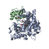 2m56C 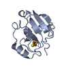 1oqrS  2zwuS S: Starting model for refinement C: citing same article ( |
|---|---|
| Similar structure data |

-Links
 PDBj
PDBj


- Assembly
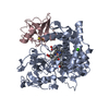
| Deposited unit | 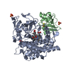
| ||||||||
|---|---|---|---|---|---|---|---|---|---|
| 1 |
| ||||||||
| Unit cell |
|
-Components
-Protein , 2 types, 2 molecules AB
| #1: Protein | Mass: 46664.973 Da / Num. of mol.: 1 / Mutation: K126C, R130C, C334A Source method: isolated from a genetically manipulated source Source: (gene. exp.) Pseudomonas putida (bacteria) / Gene: camC, cyp101 / Plasmid: pET28a / Production host: |
|---|---|
| #2: Protein | Mass: 11601.094 Da / Num. of mol.: 1 / Mutation: C73S Source method: isolated from a genetically manipulated source Source: (gene. exp.) Pseudomonas putida (bacteria) / Gene: camB / Plasmid: pET28a / Production host: |
-Non-polymers , 5 types, 42 molecules 


| #3: Chemical | ChemComp-HEM /  Mass: 616.487 Da / Num. of mol.: 1 / Source method: obtained synthetically / Formula: C34H32FeN4O4 Mass: 616.487 Da / Num. of mol.: 1 / Source method: obtained synthetically / Formula: C34H32FeN4O4 | ||||||
|---|---|---|---|---|---|---|---|
| #4: Chemical | ChemComp-SO4 /  Mass: 96.063 Da / Num. of mol.: 4 / Source method: obtained synthetically / Formula: SO4 Mass: 96.063 Da / Num. of mol.: 4 / Source method: obtained synthetically / Formula: SO4#5: Chemical | ChemComp-GOL / |  Mass: 92.094 Da / Num. of mol.: 1 / Source method: obtained synthetically / Formula: C3H8O3 Mass: 92.094 Da / Num. of mol.: 1 / Source method: obtained synthetically / Formula: C3H8O3#6: Chemical | ChemComp-FES / |  Mass: 175.820 Da / Num. of mol.: 1 / Source method: obtained synthetically / Formula: Fe2S2 Mass: 175.820 Da / Num. of mol.: 1 / Source method: obtained synthetically / Formula: Fe2S2#7: Water | ChemComp-HOH / | Mass: 18.015 Da / Num. of mol.: 35 / Source method: isolated from a natural source / Formula: H2O |
-Experimental details
-Experiment
| Experiment | Method: X-RAY DIFFRACTION / Number of used crystals: 1 |
|---|
- Sample preparation
| Crystal | Density Matthews: 2.03 Å3/Da / Density % sol: 39.5 % |
|---|---|
| Crystal grow | Temperature: 289 K / Method: vapor diffusion, hanging drop / pH: 7.5 Details: 0.1M HEPES (PH7.5), 0.1M SODIUM CHLORIDE, 1.6M AMMONIUM SULFATE, VAPOR DIFFUSION, HANGING DROP, temperature 289K |
-Data collection
| Diffraction | Mean temperature: 100 K |
|---|---|
| Diffraction source | Source: SYNCHROTRON / Site: SPring-8  / Beamline: BL44XU / Wavelength: 0.9 Å / Beamline: BL44XU / Wavelength: 0.9 Å |
| Detector | Type: RAYONIX MX225HE / Detector: CCD / Date: Feb 3, 2012 |
| Radiation | Monochromator: DOUBLE CRYSTAL SI111 / Protocol: SINGLE WAVELENGTH / Monochromatic (M) / Laue (L): M / Scattering type: x-ray |
| Radiation wavelength | Wavelength: 0.9 Å / Relative weight: 1 |
| Reflection | Resolution: 2.5→61.78 Å / Num. obs: 16251 / % possible obs: 99.6 % / Observed criterion σ(F): 1 / Observed criterion σ(I): 2 / Redundancy: 3.7 % / Rmerge(I) obs: 0.098 / Net I/σ(I): 19.4 |
| Reflection shell | Resolution: 2.5→2.54 Å / Redundancy: 3.3 % / Rmerge(I) obs: 0.44 / Mean I/σ(I) obs: 3.6 / Rsym value: 0.44 / % possible all: 98.9 |
- Processing
| Software |
| |||||||||||||||||||||||||||||||||||||||||||||||||||||||||||||||||
|---|---|---|---|---|---|---|---|---|---|---|---|---|---|---|---|---|---|---|---|---|---|---|---|---|---|---|---|---|---|---|---|---|---|---|---|---|---|---|---|---|---|---|---|---|---|---|---|---|---|---|---|---|---|---|---|---|---|---|---|---|---|---|---|---|---|---|
| Refinement | Method to determine structure: MOLECULAR REPLACEMENT Starting model: 2ZWU, 1OQR Resolution: 2.5→61.78 Å / Cor.coef. Fo:Fc: 0.942 / Cor.coef. Fo:Fc free: 0.888 / SU B: 10.585 / SU ML: 0.236 / Cross valid method: THROUGHOUT / σ(F): 0 / ESU R Free: 0.329 / Stereochemistry target values: MAXIMUM LIKELIHOOD / Details: HYDROGENS HAVE BEEN ADDED IN THE RIDING POSITIONS
| |||||||||||||||||||||||||||||||||||||||||||||||||||||||||||||||||
| Solvent computation | Ion probe radii: 0.8 Å / Shrinkage radii: 0.8 Å / VDW probe radii: 1.2 Å / Solvent model: MASK | |||||||||||||||||||||||||||||||||||||||||||||||||||||||||||||||||
| Displacement parameters | Biso mean: 31.689 Å2
| |||||||||||||||||||||||||||||||||||||||||||||||||||||||||||||||||
| Refinement step | Cycle: LAST / Resolution: 2.5→61.78 Å
| |||||||||||||||||||||||||||||||||||||||||||||||||||||||||||||||||
| Refine LS restraints |
| |||||||||||||||||||||||||||||||||||||||||||||||||||||||||||||||||
| LS refinement shell | Resolution: 2.501→2.566 Å / Total num. of bins used: 20
|
