 Movie
Movie Controller

[English] 日本語
 Yorodumi
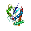
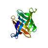
- PDB-3vtm: Structure of heme transport protein IsdH-NEAT3 from S. aureus in ... -
+ Open data

- Basic information
| Entry | Database: PDB / ID: 3vtm | ||||||
|---|---|---|---|---|---|---|---|
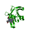
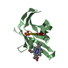
| Title | Structure of heme transport protein IsdH-NEAT3 from S. aureus in complex with Indium-porphyrin | ||||||
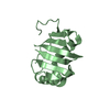
 Components Components | Iron-regulated surface determinant protein H | ||||||
 Keywords Keywords | HEME-BINDING PROTEIN / Indium / metalloporphyrin / metal selectivity / NEAT domain / Heme binding / Heme transport / Hemin / PPIX / cell wall | ||||||
| Function / homology |  Function and homology information Function and homology information | ||||||
| Biological species |   Staphylococcus aureus (bacteria) Staphylococcus aureus (bacteria) | ||||||
| Method |  X-RAY DIFFRACTION / SYNCHROTRON / MOLECULAR REPLACEMENT / molecular replacement / Resolution: 2.8 Å X-RAY DIFFRACTION / SYNCHROTRON / MOLECULAR REPLACEMENT / molecular replacement / Resolution: 2.8 Å | ||||||
 Authors Authors | Vu, N.T. / Caaveiro, J.M.M. / Moriwaki, Y. / Tsumoto, K. | ||||||
 Citation Citation | Journal: Protein Sci. / Year: 2013 Title: Selective binding of antimicrobial porphyrins to the heme-receptor IsdH-NEAT3 of Staphylococcus aureus Authors: Vu, N.T. / Moriwaki, Y. / Caaveiro, J.M.M. / Terada, T. / Tsutsumi, H. / Hamachi, I. / Shimizu, K. / Tsumoto, K. #1: Journal: Biochemistry / Year: 2011Title: Molecular basis of recognition of antibacterial porphyrins by heme-transporter IsdH-NEAT3 of Staphylococcus aureus. Authors: Moriwaki, Y. / Caaveiro, J.M. / Tanaka, Y. / Tsutsumi, H. / Hamachi, I. / Tsumoto, K. #2: Journal: J.Biol.Chem. / Year: 2008Title: Structural basis for multimeric heme complexation through a specific protein-heme interaction: the case of the third neat domain of IsdH from Staphylococcus aureus. Authors: Watanabe, M. / Tanaka, Y. / Suenaga, A. / Kuroda, M. / Yao, M. / Watanabe, N. / Arisaka, F. / Ohta, T. / Tanaka, I. / Tsumoto, K. | ||||||
| History |
|
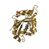
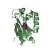
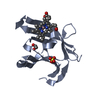
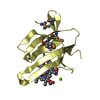
- Structure visualization
| Structure viewer | Molecule: MolmilJmol/JSmol |
|---|
- Downloads & links
-Download
| PDBx/mmCIF format | 3vtm.cif.gz | 63.1 KB | Display | PDBx/mmCIF format |
|---|---|---|---|---|
| PDB format | pdb3vtm.ent.gz | 45.6 KB | Display | PDB format |
| PDBx/mmJSON format | 3vtm.json.gz | Tree view | PDBx/mmJSON format | |
| Others |  Other downloads Other downloads |
-Validation report
| Summary document | 3vtm_validation.pdf.gz | 1.2 MB | Display | wwPDB validaton report |
|---|---|---|---|---|
| Full document | 3vtm_full_validation.pdf.gz | 1.2 MB | Display | |
| Data in XML | 3vtm_validation.xml.gz | 12.6 KB | Display | |
| Data in CIF | 3vtm_validation.cif.gz | 15.5 KB | Display | |
| Arichive directory | https://data.pdbj.org/pub/pdb/validation_reports/vt/3vtmftp://data.pdbj.org/pub/pdb/validation_reports/vt/3vtm | HTTPS FTP |
-Related structure data
| Related structure data |  3qugS S: Starting model for refinement |
|---|---|
| Similar structure data |


-Links
 PDBj
PDBj- Assembly
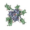
| Deposited unit | 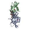
| ||||||||
|---|---|---|---|---|---|---|---|---|---|
| 1 | 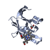
| ||||||||
| 2 | 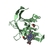
| ||||||||
| Unit cell |
|
-Components
| #1: Protein | Mass: 13003.636 Da / Num. of mol.: 2 / Fragment: NEAT domain, UNP RESIDUES 543-655 Source method: isolated from a genetically manipulated source Source: (gene. exp.) Staphylococcus aureus (bacteria) / Strain: strain Mu50 / ATCC 700699 / Gene: harA, isdH, IsdH-NEAT3, sasI, SAV1731 / Plasmid: pET28 / Production host: #2: Chemical | 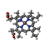  Mass: 675.460 Da / Num. of mol.: 2 / Source method: obtained synthetically / Formula: C34H32InN4O4 Mass: 675.460 Da / Num. of mol.: 2 / Source method: obtained synthetically / Formula: C34H32InN4O4#3: Chemical |   Mass: 92.094 Da / Num. of mol.: 2 / Source method: obtained synthetically / Formula: C3H8O3 Mass: 92.094 Da / Num. of mol.: 2 / Source method: obtained synthetically / Formula: C3H8O3#4: Water | ChemComp-HOH / |  Mass: 18.015 Da / Num. of mol.: 18 / Source method: isolated from a natural source / Formula: H2O Mass: 18.015 Da / Num. of mol.: 18 / Source method: isolated from a natural source / Formula: H2O |
|---|
-Experimental details
-Experiment
| Experiment | Method: X-RAY DIFFRACTION / Number of used crystals: 1 |
|---|
- Sample preparation
| Crystal | Density Matthews: 2.56 Å3/Da / Density % sol: 51.94 % / Mosaicity: 1.02 ° |
|---|---|
| Crystal grow | Temperature: 293 K / Method: vapor diffusion, hanging drop / pH: 7.3 Details: PEG-MME 3500, Sodium Iodide 0.2M, Potassium iodide 0.2M, pH 7.3, VAPOR DIFFUSION, HANGING DROP, temperature 293K |
-Data collection
| Diffraction | Mean temperature: 100 K | ||||||||||||||||||||||||||||||||||||||||||||||||||||||||||||||||||||||||||||||||||||||||||||||||||||||||||||||||||||||||||||||||||||
|---|---|---|---|---|---|---|---|---|---|---|---|---|---|---|---|---|---|---|---|---|---|---|---|---|---|---|---|---|---|---|---|---|---|---|---|---|---|---|---|---|---|---|---|---|---|---|---|---|---|---|---|---|---|---|---|---|---|---|---|---|---|---|---|---|---|---|---|---|---|---|---|---|---|---|---|---|---|---|---|---|---|---|---|---|---|---|---|---|---|---|---|---|---|---|---|---|---|---|---|---|---|---|---|---|---|---|---|---|---|---|---|---|---|---|---|---|---|---|---|---|---|---|---|---|---|---|---|---|---|---|---|---|---|
| Diffraction source | Source: SYNCHROTRON / Site: Photon Factory  / Beamline: AR-NE3A / Wavelength: 1 Å / Beamline: AR-NE3A / Wavelength: 1 Å | ||||||||||||||||||||||||||||||||||||||||||||||||||||||||||||||||||||||||||||||||||||||||||||||||||||||||||||||||||||||||||||||||||||
| Detector | Type: ADSC QUANTUM 270 / Detector: CCD / Date: Oct 23, 2011 / Details: mirrors | ||||||||||||||||||||||||||||||||||||||||||||||||||||||||||||||||||||||||||||||||||||||||||||||||||||||||||||||||||||||||||||||||||||
| Radiation | Monochromator: Numerical link type Si(111) double crystal monochromator Protocol: SINGLE WAVELENGTH / Monochromatic (M) / Laue (L): M / Scattering type: x-ray | ||||||||||||||||||||||||||||||||||||||||||||||||||||||||||||||||||||||||||||||||||||||||||||||||||||||||||||||||||||||||||||||||||||
| Radiation wavelength | Wavelength: 1 Å / Relative weight: 1 | ||||||||||||||||||||||||||||||||||||||||||||||||||||||||||||||||||||||||||||||||||||||||||||||||||||||||||||||||||||||||||||||||||||
| Reflection | Resolution: 2.8→51.822 Å / Num. all: 6932 / Num. obs: 6932 / % possible obs: 99.5 % / Observed criterion σ(F): 0 / Observed criterion σ(I): -3 / Redundancy: 4.7 % / Rsym value: 0.146 / Net I/σ(I): 8.1 | ||||||||||||||||||||||||||||||||||||||||||||||||||||||||||||||||||||||||||||||||||||||||||||||||||||||||||||||||||||||||||||||||||||
| Reflection shell | Diffraction-ID: 1
|
-Phasing
| Phasing | Method: molecular replacement | |||||||||
|---|---|---|---|---|---|---|---|---|---|---|
| Phasing MR | Model details: Phaser MODE: MR_AUTO
|
- Processing
| Software |
| |||||||||||||||||||||||||||||||||||||||||||||
|---|---|---|---|---|---|---|---|---|---|---|---|---|---|---|---|---|---|---|---|---|---|---|---|---|---|---|---|---|---|---|---|---|---|---|---|---|---|---|---|---|---|---|---|---|---|---|
| Refinement | Method to determine structure: MOLECULAR REPLACEMENT Starting model: 3QUG Resolution: 2.8→51.82 Å / Cor.coef. Fo:Fc: 0.935 / Cor.coef. Fo:Fc free: 0.891 / WRfactor Rfree: 0.2688 / WRfactor Rwork: 0.2039 / Occupancy max: 1 / Occupancy min: 1 / FOM work R set: 0.7031 / SU B: 20.159 / SU ML: 0.403 / SU Rfree: 0.454 / Cross valid method: THROUGHOUT / σ(F): 0 / ESU R Free: 0.454 / Stereochemistry target values: MAXIMUM LIKELIHOOD Details: HYDROGENS HAVE BEEN USED IF PRESENT IN THE INPUT U VALUES: REFINED INDIVIDUALLY
| |||||||||||||||||||||||||||||||||||||||||||||
| Solvent computation | Ion probe radii: 0.8 Å / Shrinkage radii: 0.8 Å / VDW probe radii: 1.2 Å / Solvent model: BABINET MODEL WITH MASK | |||||||||||||||||||||||||||||||||||||||||||||
| Displacement parameters | Biso max: 86.73 Å2 / Biso mean: 31.5934 Å2 / Biso min: 9.34 Å2
| |||||||||||||||||||||||||||||||||||||||||||||
| Refinement step | Cycle: LAST / Resolution: 2.8→51.82 Å
| |||||||||||||||||||||||||||||||||||||||||||||
| Refine LS restraints |
| |||||||||||||||||||||||||||||||||||||||||||||
| LS refinement shell | Resolution: 2.8→2.873 Å / Total num. of bins used: 20
|
