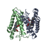
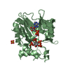
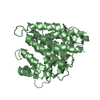
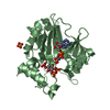
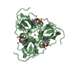
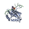
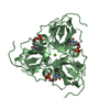
- PDB-3vrc: Crystal structure of cytochrome c' from Thermochromatium tepidum -
+
Open data
ID or keywords:
Loading...
-
Basic information
Entry
Database: PDB / ID: 3vrc
Title
Crystal structure of cytochrome c' from Thermochromatium tepidum
Components
Cytochrome c'
Keywords
ELECTRON TRANSPORT / c-type cytpchrome
Function / homology
Function and homology information
electron transport chain / electron transfer activity / periplasmic space / iron ion binding / heme binding Similarity search - Function
Cytochrome c prime, subgroup / Cytochrome c prime / Cytochrome c class II profile. / Cytochrome c, class II / Cytochrome C' / Cytochrome c/b562 / Cytochrome c/b562 / Four Helix Bundle (Hemerythrin (Met), subunit A) / Up-down Bundle / Mainly Alpha Similarity search - Domain/homology
Resolution: 1→50 Å / Cor.coef. Fo:Fc: 0.981 / Cor.coef. Fo:Fc free: 0.975 / Occupancy max: 1 / Occupancy min: 0.2 / SU B: 0.783 / SU ML: 0.018 / Cross valid method: THROUGHOUT / σ(F): 0 / ESU R: 0.022 / ESU R Free: 0.023 / Stereochemistry target values: MAXIMUM LIKELIHOOD Details: HYDROGENS HAVE BEEN USED IF PRESENT IN THE INPUT U VALUES: REFINED INDIVIDUALLY
Rfactor
Num. reflection
% reflection
Selection details
Rfree
0.1534
6350
5 %
RANDOM
Rwork
0.1292
-
-
-
obs
0.1304
121269
94.2 %
-
all
-
127619
-
-
Solvent computation
Ion probe radii: 0.8 Å / Shrinkage radii: 0.8 Å / VDW probe radii: 1.2 Å / Solvent model: BABINET MODEL WITH MASK
In the structure databanks used in Yorodumi, some data are registered as the other names, "COVID-19 virus" and "2019-nCoV". Here are the details of the virus and the list of structure data.
Jan 31, 2019. EMDB accession codes are about to change! (news from PDBe EMDB page)
EMDB accession codes are about to change! (news from PDBe EMDB page)
The allocation of 4 digits for EMDB accession codes will soon come to an end. Whilst these codes will remain in use, new EMDB accession codes will include an additional digit and will expand incrementally as the available range of codes is exhausted. The current 4-digit format prefixed with “EMD-” (i.e. EMD-XXXX) will advance to a 5-digit format (i.e. EMD-XXXXX), and so on. It is currently estimated that the 4-digit codes will be depleted around Spring 2019, at which point the 5-digit format will come into force.
The EM Navigator/Yorodumi systems omit the EMD- prefix.
Related info.:Q: What is EMD? / ID/Accession-code notation in Yorodumi/EM Navigator
Yorodumi is a browser for structure data from EMDB, PDB, SASBDB, etc.
This page is also the successor to EM Navigator detail page, and also detail information page/front-end page for Omokage search.
The word "yorodu" (or yorozu) is an old Japanese word meaning "ten thousand". "mi" (miru) is to see.
Related info.:EMDB / PDB / SASBDB / Comparison of 3 databanks / Yorodumi Search / Aug 31, 2016. New EM Navigator & Yorodumi / Yorodumi Papers / Jmol/JSmol / Function and homology information / Changes in new EM Navigator and Yorodumi
 Movie
Movie Controller


 Open data
 Basic information
 Components
 Keywords
 Function and homology information
 Thermochromatium tepidum (bacteria)
 X-RAY DIFFRACTION /
  Authors
 Citation
 Structure visualization
 Downloads & links
 Other downloads
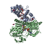


 PDBj
PDBj


 Assembly
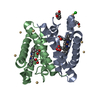


 Mass: 618.503 Da / Num. of mol.: 2 / Source method: obtained synthetically / Formula: C34H34FeN4O4
 Mass: 112.411 Da / Num. of mol.: 9 / Source method: obtained synthetically / Formula: Cd
 Mass: 35.453 Da / Num. of mol.: 1 / Source method: obtained synthetically / Formula: Cl
 Mass: 106.120 Da / Num. of mol.: 1 / Source method: obtained synthetically / Formula: C4H10O3
 Mass: 194.226 Da / Num. of mol.: 1 / Source method: obtained synthetically / Formula: C8H18O5 / Comment: precipitant*YM
 Sample preparation
 / Beamline: AR-NW12A / Wavelength: 0.8 Å
 Processing