 Movie
Movie Controller

[English] 日本語
 Yorodumi
- PDB-3vmp: Crystal structure of dextranase from Streptococcus mutans in comp... -
+ Open data

- Basic information
| Entry | Database: PDB / ID: 3vmp | ||||||
|---|---|---|---|---|---|---|---|
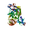
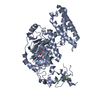
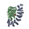
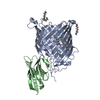
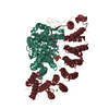
| Title | Crystal structure of dextranase from Streptococcus mutans in complex with 4,5-epoxypentyl alpha-D-glucopyranoside | ||||||
 Components Components | Dextranase | ||||||
 Keywords Keywords | HYDROLASE / TIM Barrel / immunoglobrin fold / Greek-key motif / Glycoside hydrolase family 66 | ||||||
| Function / homology |  Function and homology information Function and homology information | ||||||
| Biological species |  Streptococcus mutans (bacteria) Streptococcus mutans (bacteria) | ||||||
| Method |  X-RAY DIFFRACTION / SYNCHROTRON / MOLECULAR REPLACEMENT / Resolution: 1.88 Å X-RAY DIFFRACTION / SYNCHROTRON / MOLECULAR REPLACEMENT / Resolution: 1.88 Å | ||||||
 Authors Authors | Suzuki, N. / Fujimoto, Z. / Kim, Y.M. / Momma, M. / Okuyama, M. / Mori, H. / Funane, K. / Kimura, A. | ||||||
 Citation Citation | Journal: J.Biol.Chem. / Year: 2012 Title: Structural elucidation of dextran degradation mechanism by streptococcus mutans dextranase belonging to glycoside hydrolase family 66 Authors: Suzuki, N. / Kim, Y.M. / Fujimoto, Z. / Momma, M. / Okuyama, M. / Mori, H. / Funane, K. / Kimura, A. #1: Journal: Acta Crystallogr.,Sect.F / Year: 2011 Title: Crystallization and preliminary crystallographic analysis of dextranase from Streptococcus mutans. Authors: Suzuki, N. / Kim, Y.M. / Fujimoto, Z. / Momma, M. / Kang, H.K. / Funane, K. / Okuyama, M. / Mori, H. / Kimura, A. | ||||||
| History |
|
- Structure visualization
| Structure viewer | Molecule: MolmilJmol/JSmol |
|---|
- Downloads & links
-Download
| PDBx/mmCIF format | 3vmp.cif.gz | 146.5 KB | Display | PDBx/mmCIF format |
|---|---|---|---|---|
| PDB format | pdb3vmp.ent.gz | 111.9 KB | Display | PDB format |
| PDBx/mmJSON format | 3vmp.json.gz | Tree view | PDBx/mmJSON format | |
| Others |  Other downloads Other downloads |
-Validation report
| Arichive directory | https://data.pdbj.org/pub/pdb/validation_reports/vm/3vmpftp://data.pdbj.org/pub/pdb/validation_reports/vm/3vmp | HTTPS FTP |
|---|
-Related structure data
| Related structure data | 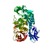 3vmnSC  3vmoC S: Starting model for refinement C: citing same article ( |
|---|---|
| Similar structure data |


-Links
 PDBj
PDBj- Assembly
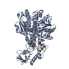
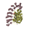
| Deposited unit | 
| ||||||||
|---|---|---|---|---|---|---|---|---|---|
| 1 |
| ||||||||
| Unit cell |
|
-Components
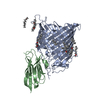
| #1: Protein | Mass: 72596.383 Da / Num. of mol.: 1 / Fragment: UNP RESIDUES 100-732 Source method: isolated from a genetically manipulated source Source: (gene. exp.) Streptococcus mutans (bacteria) / Strain: ATCC 25175 / Plasmid: pET28 / Production host: References: UniProt: F5BA50, UniProt: Q54443*PLUS, dextranase |
|---|---|
| #2: Sugar | ChemComp-E5G /   Type: D-saccharide / Mass: 266.288 Da / Num. of mol.: 1 Type: D-saccharide / Mass: 266.288 Da / Num. of mol.: 1Source method: isolated from a genetically manipulated source Formula: C11H22O7 |
| #3: Chemical | ChemComp-PO4 /   Mass: 94.971 Da / Num. of mol.: 1 / Source method: obtained synthetically / Formula: PO4 Mass: 94.971 Da / Num. of mol.: 1 / Source method: obtained synthetically / Formula: PO4 |
| #4: Water | ChemComp-HOH /  Mass: 18.015 Da / Num. of mol.: 373 / Source method: isolated from a natural source / Formula: H2O Mass: 18.015 Da / Num. of mol.: 373 / Source method: isolated from a natural source / Formula: H2O |
-Experimental details
-Experiment
| Experiment | Method: X-RAY DIFFRACTION / Number of used crystals: 1 |
|---|
- Sample preparation
| Crystal | Density Matthews: 2.03 Å3/Da / Density % sol: 39.5 % |
|---|---|
| Crystal grow | Temperature: 293 K / Method: vapor diffusion, sitting drop / pH: 4.2 Details: 30% PEGMME 2000, 0.1M phosphate-citrate, pH 4.2, VAPOR DIFFUSION, SITTING DROP, temperature 293.0K |
-Data collection
| Diffraction | Mean temperature: 95 K |
|---|---|
| Diffraction source | Source: SYNCHROTRON / Site: Photon Factory  / Beamline: BL-5A / Wavelength: 1 Å / Beamline: BL-5A / Wavelength: 1 Å |
| Detector | Type: ADSC QUANTUM 315 / Detector: CCD / Date: Feb 10, 2008 |
| Radiation | Monochromator: Si111 / Protocol: SINGLE WAVELENGTH / Monochromatic (M) / Laue (L): M / Scattering type: x-ray |
| Radiation wavelength | Wavelength: 1 Å / Relative weight: 1 |
| Reflection | Resolution: 1.88→61.41 Å / Num. obs: 45454 / % possible obs: 98.3 % / Redundancy: 6.9 % / Biso Wilson estimate: 29.4 Å2 / Rmerge(I) obs: 0.085 / Net I/σ(I): 28.9 |
| Reflection shell | Resolution: 1.9→1.97 Å / Redundancy: 5.6 % / Rmerge(I) obs: 0.267 / Mean I/σ(I) obs: 5.4 / Num. unique all: 4112 / % possible all: 89.7 |
- Processing
| Software |
| ||||||||||||||||||||||||||||||||||||||||||||||||||||||||||||||||||||||||||||||||||||||||||||||||||||||||||||||||||||||||||||||||||||||||||||||||||||||||||||||||||||||||||
|---|---|---|---|---|---|---|---|---|---|---|---|---|---|---|---|---|---|---|---|---|---|---|---|---|---|---|---|---|---|---|---|---|---|---|---|---|---|---|---|---|---|---|---|---|---|---|---|---|---|---|---|---|---|---|---|---|---|---|---|---|---|---|---|---|---|---|---|---|---|---|---|---|---|---|---|---|---|---|---|---|---|---|---|---|---|---|---|---|---|---|---|---|---|---|---|---|---|---|---|---|---|---|---|---|---|---|---|---|---|---|---|---|---|---|---|---|---|---|---|---|---|---|---|---|---|---|---|---|---|---|---|---|---|---|---|---|---|---|---|---|---|---|---|---|---|---|---|---|---|---|---|---|---|---|---|---|---|---|---|---|---|---|---|---|---|---|---|---|---|---|---|
| Refinement | Method to determine structure: MOLECULAR REPLACEMENT Starting model: 3VMN Resolution: 1.88→50 Å / Cor.coef. Fo:Fc: 0.945 / Cor.coef. Fo:Fc free: 0.923 / SU B: 3.851 / SU ML: 0.114 / Cross valid method: THROUGHOUT / ESU R Free: 0.163 / Stereochemistry target values: MAXIMUM LIKELIHOOD / Details: HYDROGENS HAVE BEEN ADDED IN THE RIDING POSITIONS
| ||||||||||||||||||||||||||||||||||||||||||||||||||||||||||||||||||||||||||||||||||||||||||||||||||||||||||||||||||||||||||||||||||||||||||||||||||||||||||||||||||||||||||
| Solvent computation | Ion probe radii: 0.8 Å / Shrinkage radii: 0.8 Å / VDW probe radii: 1.4 Å / Solvent model: MASK | ||||||||||||||||||||||||||||||||||||||||||||||||||||||||||||||||||||||||||||||||||||||||||||||||||||||||||||||||||||||||||||||||||||||||||||||||||||||||||||||||||||||||||
| Displacement parameters | Biso mean: 26.537 Å2
| ||||||||||||||||||||||||||||||||||||||||||||||||||||||||||||||||||||||||||||||||||||||||||||||||||||||||||||||||||||||||||||||||||||||||||||||||||||||||||||||||||||||||||
| Refinement step | Cycle: LAST / Resolution: 1.88→50 Å
| ||||||||||||||||||||||||||||||||||||||||||||||||||||||||||||||||||||||||||||||||||||||||||||||||||||||||||||||||||||||||||||||||||||||||||||||||||||||||||||||||||||||||||
| Refine LS restraints |
| ||||||||||||||||||||||||||||||||||||||||||||||||||||||||||||||||||||||||||||||||||||||||||||||||||||||||||||||||||||||||||||||||||||||||||||||||||||||||||||||||||||||||||
| LS refinement shell | Resolution: 1.884→1.933 Å / Total num. of bins used: 20
|
