 Movie
Movie Controller

[English] 日本語
 Yorodumi
- PDB-6qgy: Crystal structure of E.coli BamA beta-barrel in complex with nano... -
+ Open data

- Basic information
| Entry | Database: PDB / ID: 6qgy | ||||||
|---|---|---|---|---|---|---|---|
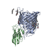
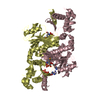
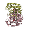
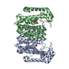
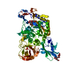
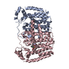
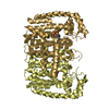
| Title | Crystal structure of E.coli BamA beta-barrel in complex with nanobody B12 | ||||||
 Components Components |
| ||||||
 Keywords Keywords | MEMBRANE PROTEIN / Beta-Barrel / outer membrane / protein insertion / protein folding / protein maturation | ||||||
| Function / homology |  Function and homology information Function and homology informationBam protein complex / Gram-negative-bacterium-type cell outer membrane assembly / protein insertion into membrane Similarity search - Function | ||||||
| Biological species |   | ||||||
| Method |  X-RAY DIFFRACTION / SYNCHROTRON / MOLECULAR REPLACEMENT / Resolution: 2.509 Å X-RAY DIFFRACTION / SYNCHROTRON / MOLECULAR REPLACEMENT / Resolution: 2.509 Å | ||||||
 Authors Authors | Hartmann, J.-B. / Kaur, H. / Jakob, R.P. / Zahn, M. / Zimmermann, I. / Seeger, M. / Maier, T. / Hiller, S. | ||||||
 Citation Citation | Journal: J.Biomol.Nmr / Year: 2019 Title: Identification of conformation-selective nanobodies against the membrane protein insertase BamA by an integrated structural biology approach. Authors: Kaur, H. / Hartmann, J.B. / Jakob, R.P. / Zahn, M. / Zimmermann, I. / Maier, T. / Seeger, M.A. / Hiller, S. | ||||||
| History |
|
- Structure visualization
| Structure viewer | Molecule: MolmilJmol/JSmol |
|---|
- Downloads & links
-Download
| PDBx/mmCIF format | 6qgy.cif.gz | 373.7 KB | Display | PDBx/mmCIF format |
|---|---|---|---|---|
| PDB format | pdb6qgy.ent.gz | 308 KB | Display | PDB format |
| PDBx/mmJSON format | 6qgy.json.gz | Tree view | PDBx/mmJSON format | |
| Others |  Other downloads Other downloads |
-Validation report
| Arichive directory | https://data.pdbj.org/pub/pdb/validation_reports/qg/6qgyftp://data.pdbj.org/pub/pdb/validation_reports/qg/6qgy | HTTPS FTP |
|---|
-Related structure data
| Related structure data |  6qgwC  6qgxC  4n75S S: Starting model for refinement C: citing same article ( |
|---|---|
| Similar structure data |


-Links
 PDBj
PDBj

- Assembly
| Deposited unit | 
| ||||||||
|---|---|---|---|---|---|---|---|---|---|
| 1 | 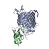
| ||||||||
| 2 | 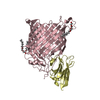
| ||||||||
| Unit cell |
| ||||||||
| Components on special symmetry positions |
|
-Components
| #1: Protein | Mass: 46022.789 Da / Num. of mol.: 2 Source method: isolated from a genetically manipulated source Source: (gene. exp.) #2: Antibody | Mass: 13637.087 Da / Num. of mol.: 2 Source method: isolated from a genetically manipulated source Source: (gene. exp.) #3: Chemical | ChemComp-C8E / (   Mass: 306.438 Da / Num. of mol.: 10 / Source method: obtained synthetically / Formula: C16H34O5 / Comment: C8E, detergent*YM Mass: 306.438 Da / Num. of mol.: 10 / Source method: obtained synthetically / Formula: C16H34O5 / Comment: C8E, detergent*YM#4: Water | ChemComp-HOH / |  Mass: 18.015 Da / Num. of mol.: 317 / Source method: isolated from a natural source / Formula: H2O Mass: 18.015 Da / Num. of mol.: 317 / Source method: isolated from a natural source / Formula: H2OHas protein modification | Y | |
|---|
-Experimental details
-Experiment
| Experiment | Method: X-RAY DIFFRACTION / Number of used crystals: 1 |
|---|
- Sample preparation
| Crystal | Density Matthews: 4.37 Å3/Da / Density % sol: 71.84 % |
|---|---|
| Crystal grow | Temperature: 293 K / Method: vapor diffusion, hanging drop Details: 32% PEG 400, 0.2 M ammonium phosphate, 0.1 M ammonium sulfate, 0.1 M sodium citrate pH 4.5 |
-Data collection
| Diffraction | Mean temperature: 100 K / Serial crystal experiment: N |
|---|---|
| Diffraction source | Source: SYNCHROTRON / Site: SLS  / Beamline: X06SA / Wavelength: 0.99998 Å / Beamline: X06SA / Wavelength: 0.99998 Å |
| Detector | Type: DECTRIS EIGER X 16M / Detector: PIXEL / Date: Feb 2, 2018 |
| Radiation | Protocol: SINGLE WAVELENGTH / Monochromatic (M) / Laue (L): M / Scattering type: x-ray |
| Radiation wavelength | Wavelength: 0.99998 Å / Relative weight: 1 |
| Reflection | Resolution: 2.509→48.484 Å / Num. obs: 69491 / % possible obs: 98.5 % / Redundancy: 5.8 % / CC1/2: 0.998 / Rmerge(I) obs: 0.073 / Rpim(I) all: 0.051 / Net I/σ(I): 11.9 |
| Reflection shell | Resolution: 2.51→2.57 Å / Redundancy: 5.3 % / Rmerge(I) obs: 0.973 / Mean I/σ(I) obs: 1.2 / Num. unique obs: 3901 / CC1/2: 0.621 / Rpim(I) all: 0.699 / % possible all: 86.3 |
- Processing
| Software |
| ||||||||||||||||||||||||||||||||||||||||||||||||||||||||||||||||||||||||||||||||||||||||||||||||||||||||||||||||||||||||||||||||||||||||||||||||||||||||||||||||||||||||||||||||||||||
|---|---|---|---|---|---|---|---|---|---|---|---|---|---|---|---|---|---|---|---|---|---|---|---|---|---|---|---|---|---|---|---|---|---|---|---|---|---|---|---|---|---|---|---|---|---|---|---|---|---|---|---|---|---|---|---|---|---|---|---|---|---|---|---|---|---|---|---|---|---|---|---|---|---|---|---|---|---|---|---|---|---|---|---|---|---|---|---|---|---|---|---|---|---|---|---|---|---|---|---|---|---|---|---|---|---|---|---|---|---|---|---|---|---|---|---|---|---|---|---|---|---|---|---|---|---|---|---|---|---|---|---|---|---|---|---|---|---|---|---|---|---|---|---|---|---|---|---|---|---|---|---|---|---|---|---|---|---|---|---|---|---|---|---|---|---|---|---|---|---|---|---|---|---|---|---|---|---|---|---|---|---|---|---|
| Refinement | Method to determine structure: MOLECULAR REPLACEMENT Starting model: 4n75 Resolution: 2.509→48.484 Å / SU ML: 0.38 / Cross valid method: FREE R-VALUE / σ(F): 1.33 / Phase error: 28.64
| ||||||||||||||||||||||||||||||||||||||||||||||||||||||||||||||||||||||||||||||||||||||||||||||||||||||||||||||||||||||||||||||||||||||||||||||||||||||||||||||||||||||||||||||||||||||
| Solvent computation | Shrinkage radii: 0.9 Å / VDW probe radii: 1.11 Å | ||||||||||||||||||||||||||||||||||||||||||||||||||||||||||||||||||||||||||||||||||||||||||||||||||||||||||||||||||||||||||||||||||||||||||||||||||||||||||||||||||||||||||||||||||||||
| Refinement step | Cycle: LAST / Resolution: 2.509→48.484 Å
| ||||||||||||||||||||||||||||||||||||||||||||||||||||||||||||||||||||||||||||||||||||||||||||||||||||||||||||||||||||||||||||||||||||||||||||||||||||||||||||||||||||||||||||||||||||||
| Refine LS restraints |
| ||||||||||||||||||||||||||||||||||||||||||||||||||||||||||||||||||||||||||||||||||||||||||||||||||||||||||||||||||||||||||||||||||||||||||||||||||||||||||||||||||||||||||||||||||||||
| LS refinement shell |
|
