Resolution: 1.98→2.07 Å / Redundancy: 2.6 % / Rmerge(I) obs: 0.84 / Mean I/σ(I) obs: 0.61 / % possible all: 98.4
-
Processing
Software
Name
Version
Classification
ADSC
Quantum
datacollection
CNS
refinement
HKL-2000
datareduction
HKL-2000
datascaling
CNS
phasing
Refinement
Method to determine structure: MOLECULAR REPLACEMENT / Resolution: 2→48.36 Å / Rfactor Rfree error: 0.004 / Occupancy max: 1 / Occupancy min: 0.74 / Data cutoff high absF: 2910630 / Data cutoff low absF: 0 / Isotropic thermal model: RESTRAINED / Cross valid method: THROUGHOUT / σ(F): 0 Details: FOR STRONG INHIBITORS (SHA), THE HIGHER B-FACTOR AND LOWER OCCUPANCY IN SER305ALA VARIANT CORRELATED WITH THE HIGHER BINDING ENERGIES OF CHAIN B AS COMPARED WITH CHAIN A, ESPECIALLY AT LOW ...Details: FOR STRONG INHIBITORS (SHA), THE HIGHER B-FACTOR AND LOWER OCCUPANCY IN SER305ALA VARIANT CORRELATED WITH THE HIGHER BINDING ENERGIES OF CHAIN B AS COMPARED WITH CHAIN A, ESPECIALLY AT LOW SHA LIGAND CONCENTRATION.
In the structure databanks used in Yorodumi, some data are registered as the other names, "COVID-19 virus" and "2019-nCoV". Here are the details of the virus and the list of structure data.
Jan 31, 2019. EMDB accession codes are about to change! (news from PDBe EMDB page)
EMDB accession codes are about to change! (news from PDBe EMDB page)
The allocation of 4 digits for EMDB accession codes will soon come to an end. Whilst these codes will remain in use, new EMDB accession codes will include an additional digit and will expand incrementally as the available range of codes is exhausted. The current 4-digit format prefixed with “EMD-” (i.e. EMD-XXXX) will advance to a 5-digit format (i.e. EMD-XXXXX), and so on. It is currently estimated that the 4-digit codes will be depleted around Spring 2019, at which point the 5-digit format will come into force.
The EM Navigator/Yorodumi systems omit the EMD- prefix.
Related info.:Q: What is EMD? / ID/Accession-code notation in Yorodumi/EM Navigator
Yorodumi is a browser for structure data from EMDB, PDB, SASBDB, etc.
This page is also the successor to EM Navigator detail page, and also detail information page/front-end page for Omokage search.
The word "yorodu" (or yorozu) is an old Japanese word meaning "ten thousand". "mi" (miru) is to see.
Related info.:EMDB / PDB / SASBDB / Comparison of 3 databanks / Yorodumi Search / Aug 31, 2016. New EM Navigator & Yorodumi / Yorodumi Papers / Jmol/JSmol / Function and homology information / Changes in new EM Navigator and Yorodumi
 Movie
Movie Controller

 Yorodumi
 Open data
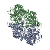
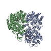
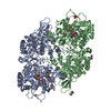
 Basic information
 Components
 Keywords
 Function and homology information
 Haloarcula marismortui (Halophile)
 X-RAY DIFFRACTION /
  Authors
 Citation
 Structure visualization
 Downloads & links
 Other downloads


 PDBj
PDBj
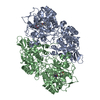
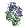
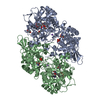
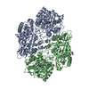
 Assembly

 Mass: 616.487 Da / Num. of mol.: 2 / Source method: obtained synthetically / Formula: C34H32FeN4O4
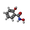
 Mass: 153.135 Da / Num. of mol.: 2 / Source method: obtained synthetically / Formula: C7H7NO3
 Mass: 18.015 Da / Num. of mol.: 242 / Source method: isolated from a natural source / Formula: H2O
 Sample preparation
 / Beamline: BL-17A / Wavelength: 1 Å
 Processing