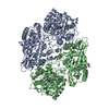
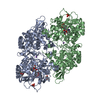
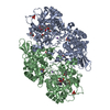
[English] 日本語
Yorodumi
- PDB-5cjh: Crystal Structure of Eukaryotic Oxoiron MagKatG2 at pH 8.5 -

+
Open data


ID or keywords:

Loading...

-
Basic information

Entry
Database: PDB / ID: 5cjh
TitleCrystal Structure of Eukaryotic Oxoiron MagKatG2 at pH 8.5
ComponentsCatalase-peroxidase 2
KeywordsOXIDOREDUCTASE / COMPOUND I / OXOIRON CATALASE-PEROXIDASE
Function / homology
Function and homology information


catalase-peroxidase / catalase activity / hydrogen peroxide catabolic process / response to oxidative stress / heme binding / extracellular region / metal ion binding
Similarity search - Function
Catalase-peroxidase haem / Peroxidase; domain 2 / Peroxidase, domain 2 / Peroxidase; domain 1 - #10 / Peroxidase; domain 1 / Peroxidases heam-ligand binding site / Peroxidase, active site / Peroxidases active site signature. / Plant heme peroxidase family profile. / Haem peroxidase ...Catalase-peroxidase haem / Peroxidase; domain 2 / Peroxidase, domain 2 / Peroxidase; domain 1 - #10 / Peroxidase; domain 1 / Peroxidases heam-ligand binding site / Peroxidase, active site / Peroxidases active site signature. / Plant heme peroxidase family profile. / Haem peroxidase / Peroxidase / Peroxidases proximal heme-ligand signature. / Haem peroxidase superfamily / Orthogonal Bundle / Mainly Alpha
Similarity search - Domain/homology
Peroxidized Heme / HYDROXIDE ION / Catalase-peroxidase 2
Similarity search - Component
Biological speciesMagnaporthe oryzae (rice blast fungus)
MethodX-RAY DIFFRACTION / SYNCHROTRON / MOLECULAR REPLACEMENT / Resolution: 1.6 Å
AuthorsGasselhuber, B. / Obinger, C. / Fita, I. / Carpena, X.
CitationJournal: Biochemistry / Year: 2015
Title: Eukaryotic Catalase-Peroxidase: The Role of the Trp-Tyr-Met Adduct in Protein Stability, Substrate Accessibility, and Catalysis of Hydrogen Peroxide Dismutation.
Authors: Gasselhuber, B. / Carpena, X. / Graf, M.M. / Pirker, K.F. / Nicolussi, A. / Sundermann, A. / Hofbauer, S. / Zamocky, M. / Furtmuller, P.G. / Jakopitsch, C. / Oostenbrink, C. / Fita, I. / Obinger, C.
History
DepositionJul 14, 2015Deposition site: RCSB / Processing site: PDBE
Revision 1.0Sep 2, 2015Provider: repository / Type: Initial release
Revision 1.1Sep 16, 2015Group: Database references
Revision 1.2Oct 28, 2015Group: Structure summary
Revision 1.3Feb 20, 2019Group: Advisory / Data collection / Derived calculations
Category: pdbx_data_processing_status / pdbx_unobs_or_zero_occ_atoms ...pdbx_data_processing_status / pdbx_unobs_or_zero_occ_atoms / pdbx_validate_close_contact / struct_conn / struct_conn_type
Revision 1.4Jan 10, 2024Group: Advisory / Data collection ...Advisory / Data collection / Database references / Derived calculations / Refinement description
Category: chem_comp_atom / chem_comp_bond ...chem_comp_atom / chem_comp_bond / database_2 / pdbx_initial_refinement_model / pdbx_unobs_or_zero_occ_atoms / struct_conn
Item: _database_2.pdbx_DOI / _database_2.pdbx_database_accession ..._database_2.pdbx_DOI / _database_2.pdbx_database_accession / _struct_conn.pdbx_dist_value / _struct_conn.ptnr1_auth_asym_id / _struct_conn.ptnr1_auth_comp_id / _struct_conn.ptnr1_auth_seq_id / _struct_conn.ptnr1_label_asym_id / _struct_conn.ptnr1_label_atom_id / _struct_conn.ptnr1_label_comp_id / _struct_conn.ptnr1_label_seq_id / _struct_conn.ptnr2_auth_asym_id / _struct_conn.ptnr2_auth_comp_id / _struct_conn.ptnr2_auth_seq_id / _struct_conn.ptnr2_label_asym_id / _struct_conn.ptnr2_label_atom_id / _struct_conn.ptnr2_label_comp_id

-
Structure visualization

Structure viewerMolecule:
MolmilJmol/JSmol

Downloads & links

-
Assembly

Deposited unit
A: Catalase-peroxidase 2
B: Catalase-peroxidase 2
hetero molecules


Theoretical massNumber of molelcules
Total (without water)168,2556
Polymers166,9202
Non-polymers1,3354
Water28,3381573
1


  • Idetical with deposited unit
  • defined by software
TypeNameSymmetry operationNumber
identity operation1_555x,y,z1
Buried area9830 Å2
ΔGint-54 kcal/mol
Surface area50530 Å2
MethodPISA
Unit cell
Length a, b, c (Å)103.530, 109.540, 132.150
Angle α, β, γ (deg.)90.00, 90.00, 90.00
Int Tables number19
Space group name H-MP212121

-
Components

#1: Protein Catalase-peroxidase 2 / / CP 2 / Peroxidase/catalase 2


Mass: 83460.188 Da / Num. of mol.: 2 / Fragment: RESIDUES 24-786
Source method: isolated from a genetically manipulated source
Source: (gene. exp.) Magnaporthe oryzae (strain 70-15 / ATCC MYA-4617 / FGSC 8958) (fungus)
Gene: KATG2, CPXB, MagKatG2, MGG_09834 / Production host: Escherichia coli (E. coli) / References: UniProt: A4QUT2, catalase-peroxidase
#2: Chemical ChemComp-522 / Peroxidized Heme


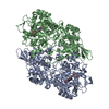
Mass: 650.502 Da / Num. of mol.: 2 / Source method: obtained synthetically / Formula: C34H34FeN4O6
#3: Chemical ChemComp-OH / HYDROXIDE ION / Hydroxide


Mass: 17.007 Da / Num. of mol.: 2 / Source method: obtained synthetically / Formula: HO
#4: Water ChemComp-HOH / water / Water


Mass: 18.015 Da / Num. of mol.: 1573 / Source method: isolated from a natural source / Formula: H2O

-
Experimental details

-
Experiment

ExperimentMethod: X-RAY DIFFRACTION

-
Sample preparation

CrystalDensity Matthews: 2.29 Å3/Da / Density % sol: 46.3 %
Crystal growTemperature: 277 K / Method: vapor diffusion, hanging drop / pH: 4.6
Details: 15% PEG4000, 0.1 M sodium acetate, pH 4.6 (+ PAA soaking + pH 8.5 soaking)

-
Data collection

DiffractionMean temperature: 100 K
Diffraction sourceSource: SYNCHROTRON / Site: ESRF / Beamline: ID23-1 / Wavelength: 0.9717 Å
DetectorType: DECTRIS PILATUS 6M / Detector: PIXEL / Date: Jul 14, 2013
RadiationMonochromator: Si (111) / Protocol: SINGLE WAVELENGTH / Monochromatic (M) / Laue (L): M / Scattering type: x-ray
Radiation wavelengthWavelength: 0.9717 Å / Relative weight: 1
ReflectionResolution: 1.6→20 Å / Num. obs: 196582 / % possible obs: 99.5 % / Redundancy: 4.5 % / Rmerge(I) obs: 0.099 / Rsym value: 0.087 / Net I/σ(I): 11.2
Reflection shellResolution: 1.6→1.64 Å / Redundancy: 4.6 % / Rmerge(I) obs: 0.73 / Mean I/σ(I) obs: 2.4 / % possible all: 99.3

-
Processing

Software
NameVersionClassification
REFMAC5.7.0029refinement
XDSdata reduction
XSCALEdata scaling
MOLREPphasing
RefinementMethod to determine structure: MOLECULAR REPLACEMENT
Starting model: 3UT2
Resolution: 1.6→19.94 Å / Cor.coef. Fo:Fc: 0.965 / Cor.coef. Fo:Fc free: 0.955 / SU B: 1.51 / SU ML: 0.053 / Cross valid method: THROUGHOUT / ESU R: 0.084 / ESU R Free: 0.082 / Stereochemistry target values: MAXIMUM LIKELIHOOD / Details: HYDROGENS HAVE BEEN ADDED IN THE RIDING POSITIONS
RfactorNum. reflection% reflectionSelection details
Rfree0.18992 9850 5 %RANDOM
Rwork0.16472 ---
obs0.16597 186717 99.44 %-
Solvent computationIon probe radii: 0.8 Å / Shrinkage radii: 0.8 Å / VDW probe radii: 1.2 Å / Solvent model: MASK
Displacement parametersBiso mean: 17.96 Å2
Baniso -1Baniso -2Baniso -3
1--0.06 Å20 Å20 Å2
2---0.5 Å2-0 Å2
3---0.56 Å2
Refinement stepCycle: LAST / Resolution: 1.6→19.94 Å
ProteinNucleic acidLigandSolventTotal
Num. atoms11283 0 92 1573 12948
Refine LS restraints
Refine-IDTypeDev idealDev ideal targetNumber
X-RAY DIFFRACTIONr_bond_refined_d0.0080.01911853
X-RAY DIFFRACTIONr_bond_other_d0.0010.0210918
X-RAY DIFFRACTIONr_angle_refined_deg1.3941.94916172
X-RAY DIFFRACTIONr_angle_other_deg0.794325124
X-RAY DIFFRACTIONr_dihedral_angle_1_deg5.33251529
X-RAY DIFFRACTIONr_dihedral_angle_2_deg35.31824.549565
X-RAY DIFFRACTIONr_dihedral_angle_3_deg12.12151878
X-RAY DIFFRACTIONr_dihedral_angle_4_deg15.2961574
X-RAY DIFFRACTIONr_chiral_restr0.0780.21694
X-RAY DIFFRACTIONr_gen_planes_refined0.0060.02113936
X-RAY DIFFRACTIONr_gen_planes_other0.0050.022828
X-RAY DIFFRACTIONr_nbd_refined
X-RAY DIFFRACTIONr_nbd_other
X-RAY DIFFRACTIONr_nbtor_refined
X-RAY DIFFRACTIONr_nbtor_other
X-RAY DIFFRACTIONr_xyhbond_nbd_refined
X-RAY DIFFRACTIONr_xyhbond_nbd_other
X-RAY DIFFRACTIONr_metal_ion_refined
X-RAY DIFFRACTIONr_metal_ion_other
X-RAY DIFFRACTIONr_symmetry_vdw_refined
X-RAY DIFFRACTIONr_symmetry_vdw_other
X-RAY DIFFRACTIONr_symmetry_hbond_refined
X-RAY DIFFRACTIONr_symmetry_hbond_other
X-RAY DIFFRACTIONr_symmetry_metal_ion_refined
X-RAY DIFFRACTIONr_symmetry_metal_ion_other
X-RAY DIFFRACTIONr_mcbond_it
X-RAY DIFFRACTIONr_mcbond_other
X-RAY DIFFRACTIONr_mcangle_it
X-RAY DIFFRACTIONr_mcangle_other
X-RAY DIFFRACTIONr_scbond_it
X-RAY DIFFRACTIONr_scbond_other
X-RAY DIFFRACTIONr_scangle_it
X-RAY DIFFRACTIONr_scangle_other
X-RAY DIFFRACTIONr_long_range_B_refined
X-RAY DIFFRACTIONr_long_range_B_other
X-RAY DIFFRACTIONr_rigid_bond_restr
X-RAY DIFFRACTIONr_sphericity_free
X-RAY DIFFRACTIONr_sphericity_bonded
LS refinement shellResolution: 1.6→1.641 Å / Total num. of bins used: 20
RfactorNum. reflection% reflection
Rfree0.29 759 -
Rwork0.259 13557 -
obs--99.31 %

+
About Yorodumi

-
News

-
Feb 9, 2022. New format data for meta-information of EMDB entries

New format data for meta-information of EMDB entries

  • Version 3 of the EMDB header file is now the official format.
  • The previous official version 1.9 will be removed from the archive.

Related info.:EMDB header

External links:wwPDB to switch to version 3 of the EMDB data model

-
Aug 12, 2020. Covid-19 info

Covid-19 info

URL: https://pdbj.org/emnavi/covid19.php

New page: Covid-19 featured information page in EM Navigator.

Related info.:Covid-19 info / Mar 5, 2020. Novel coronavirus structure data

+
Mar 5, 2020. Novel coronavirus structure data

Novel coronavirus structure data

Related info.:Yorodumi Speices / Aug 12, 2020. Covid-19 info

External links:COVID-19 featured content - PDBj / Molecule of the Month (242):Coronavirus Proteases

+
Jan 31, 2019. EMDB accession codes are about to change! (news from PDBe EMDB page)

EMDB accession codes are about to change! (news from PDBe EMDB page)

  • The allocation of 4 digits for EMDB accession codes will soon come to an end. Whilst these codes will remain in use, new EMDB accession codes will include an additional digit and will expand incrementally as the available range of codes is exhausted. The current 4-digit format prefixed with “EMD-” (i.e. EMD-XXXX) will advance to a 5-digit format (i.e. EMD-XXXXX), and so on. It is currently estimated that the 4-digit codes will be depleted around Spring 2019, at which point the 5-digit format will come into force.
  • The EM Navigator/Yorodumi systems omit the EMD- prefix.

Related info.:Q: What is EMD? / ID/Accession-code notation in Yorodumi/EM Navigator

External links:EMDB Accession Codes are Changing Soon! / Contact to PDBj

+
Jul 12, 2017. Major update of PDB

Major update of PDB

  • wwPDB released updated PDB data conforming to the new PDBx/mmCIF dictionary.
  • This is a major update changing the version number from 4 to 5, and with Remediation, in which all the entries are updated.
  • In this update, many items about electron microscopy experimental information are reorganized (e.g. em_software).
  • Now, EM Navigator and Yorodumi are based on the updated data.

External links:wwPDB Remediation / Enriched Model Files Conforming to OneDep Data Standards Now Available in the PDB FTP Archive

-
Yorodumi

Thousand views of thousand structures

  • Yorodumi is a browser for structure data from EMDB, PDB, SASBDB, etc.
  • This page is also the successor to EM Navigator detail page, and also detail information page/front-end page for Omokage search.
  • The word "yorodu" (or yorozu) is an old Japanese word meaning "ten thousand". "mi" (miru) is to see.

Related info.:EMDB / PDB / SASBDB / Comparison of 3 databanks / Yorodumi Search / Aug 31, 2016. New EM Navigator & Yorodumi / Yorodumi Papers / Jmol/JSmol / Function and homology information / Changes in new EM Navigator and Yorodumi

Read more