 Movie
Movie Controller

[English] 日本語
 Yorodumi
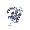
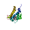
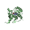
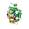
- PDB-3vgp: Crystal structure of the C-terminal globular domain of oligosacch... -
+ Open data

- Basic information
| Entry | Database: PDB / ID: 3vgp | ||||||
|---|---|---|---|---|---|---|---|
| Title | Crystal structure of the C-terminal globular domain of oligosaccharyltransferase (AF_0329) from Archaeoglobus fulgidus | ||||||
 Components Components | Transmembrane oligosaccharyl transferase, putative | ||||||
 Keywords Keywords | TRANSFERASE / glycosyltransferase / cell membrane | ||||||
| Function / homology |  Function and homology information Function and homology informationdolichyl-phosphooligosaccharide-protein glycotransferase / oligosaccharyl transferase activity / metal ion binding / plasma membrane Similarity search - Function | ||||||
| Biological species |   Archaeoglobus fulgidus (archaea) Archaeoglobus fulgidus (archaea) | ||||||
| Method |  X-RAY DIFFRACTION / SYNCHROTRON / SAD / Resolution: 1.75 Å X-RAY DIFFRACTION / SYNCHROTRON / SAD / Resolution: 1.75 Å | ||||||
 Authors Authors | Matsumoto, S. / Igura, M. / Nyirenda, J. / Yuzawa, S. / Noda, N.N. / Inagaki, F. / Kohda, D. | ||||||
 Citation Citation | Journal: Biochemistry / Year: 2012 Title: Crystal Structure of the C-Terminal Globular Domain of Oligosaccharyltransferase from Archaeoglobus fulgidus at 1.75 A Resolution Authors: Matsumoto, S. / Igura, M. / Nyirenda, J. / Matsumoto, M. / Yuzawa, S. / Noda, N.N. / Inagaki, F. / Kohda, D. | ||||||
| History |
|
- Structure visualization
| Structure viewer | Molecule: MolmilJmol/JSmol |
|---|
- Downloads & links
-Download
| PDBx/mmCIF format | 3vgp.cif.gz | 48.6 KB | Display | PDBx/mmCIF format |
|---|---|---|---|---|
| PDB format | pdb3vgp.ent.gz | 34.1 KB | Display | PDB format |
| PDBx/mmJSON format | 3vgp.json.gz | Tree view | PDBx/mmJSON format | |
| Others |  Other downloads Other downloads |
-Validation report
| Arichive directory | https://data.pdbj.org/pub/pdb/validation_reports/vg/3vgpftp://data.pdbj.org/pub/pdb/validation_reports/vg/3vgp | HTTPS FTP |
|---|
-Related structure data
| Similar structure data |
|---|


-Links
 PDBj
PDBj
- Assembly
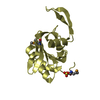
| Deposited unit | 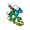
| ||||||||
|---|---|---|---|---|---|---|---|---|---|
| 1 |
| ||||||||
| Unit cell |
|
-Components
| #1: Protein | Mass: 19292.887 Da / Num. of mol.: 1 / Fragment: C-terminal globular domain / Mutation: C482A Source method: isolated from a genetically manipulated source Source: (gene. exp.) Archaeoglobus fulgidus (archaea) / Strain: DSM 4304 / Gene: AF_0329 / Plasmid: pET-47b / Production host:  |
|---|---|
| #2: Water | ChemComp-HOH /  Mass: 18.015 Da / Num. of mol.: 154 / Source method: isolated from a natural source / Formula: H2O Mass: 18.015 Da / Num. of mol.: 154 / Source method: isolated from a natural source / Formula: H2O |
| Has protein modification | Y |
-Experimental details
-Experiment
| Experiment | Method: X-RAY DIFFRACTION / Number of used crystals: 1 |
|---|
- Sample preparation
| Crystal | Density Matthews: 2.29 Å3/Da / Density % sol: 46.33 % |
|---|---|
| Crystal grow | Temperature: 293 K / Method: vapor diffusion, hanging drop / pH: 6.5 Details: 12.5% PEG 3350, 0.1M bis-Tris, pH 6.5, VAPOR DIFFUSION, HANGING DROP, temperature 293K |
-Data collection
| Diffraction source | Source: SYNCHROTRON / Site: SPring-8  / Beamline: BL44XU / Wavelength: 0.9798 Å / Beamline: BL44XU / Wavelength: 0.9798 Å |
|---|---|
| Detector | Type: BRUKER SMART 6500 / Detector: CCD / Date: Jul 22, 2010 |
| Radiation | Monochromator: a double-crystal monochromator and a horizontal focusing mirror Protocol: SINGLE WAVELENGTH / Monochromatic (M) / Laue (L): M / Scattering type: x-ray |
| Radiation wavelength | Wavelength: 0.9798 Å / Relative weight: 1 |
| Reflection | Resolution: 1.75→50 Å / Num. all: 19197 / Num. obs: 18986 / % possible obs: 98.9 % / Observed criterion σ(F): 0 / Observed criterion σ(I): 3 / Redundancy: 27 % / Biso Wilson estimate: 23 Å2 / Rmerge(I) obs: 0.062 / Net I/σ(I): 98.6 |
| Reflection shell | Resolution: 1.75→1.78 Å / Redundancy: 24.8 % / Rmerge(I) obs: 0.399 / Mean I/σ(I) obs: 13 / Num. unique all: 853 / % possible all: 93.9 |
- Processing
| Software |
| ||||||||||||||||||||||||||||||||||||||||||||||||||||||||||||||||||||||||||||||||
|---|---|---|---|---|---|---|---|---|---|---|---|---|---|---|---|---|---|---|---|---|---|---|---|---|---|---|---|---|---|---|---|---|---|---|---|---|---|---|---|---|---|---|---|---|---|---|---|---|---|---|---|---|---|---|---|---|---|---|---|---|---|---|---|---|---|---|---|---|---|---|---|---|---|---|---|---|---|---|---|---|---|
| Refinement | Method to determine structure: SAD / Resolution: 1.75→40.54 Å / Rfactor Rfree error: 0.006 / Data cutoff high absF: 234062.63 / Data cutoff low absF: 0 / Isotropic thermal model: RESTRAINED / Cross valid method: THROUGHOUT / σ(F): 0 / Stereochemistry target values: Engh & Huber / Details: BULK SOLVENT MODEL USED
| ||||||||||||||||||||||||||||||||||||||||||||||||||||||||||||||||||||||||||||||||
| Solvent computation | Solvent model: FLAT MODEL / Bsol: 55.7233 Å2 / ksol: 0.45 e/Å3 | ||||||||||||||||||||||||||||||||||||||||||||||||||||||||||||||||||||||||||||||||
| Displacement parameters | Biso mean: 26.6 Å2
| ||||||||||||||||||||||||||||||||||||||||||||||||||||||||||||||||||||||||||||||||
| Refine analyze |
| ||||||||||||||||||||||||||||||||||||||||||||||||||||||||||||||||||||||||||||||||
| Refinement step | Cycle: LAST / Resolution: 1.75→40.54 Å
| ||||||||||||||||||||||||||||||||||||||||||||||||||||||||||||||||||||||||||||||||
| Refine LS restraints |
| ||||||||||||||||||||||||||||||||||||||||||||||||||||||||||||||||||||||||||||||||
| LS refinement shell | Resolution: 1.75→1.86 Å / Rfactor Rfree error: 0.018 / Total num. of bins used: 6
| ||||||||||||||||||||||||||||||||||||||||||||||||||||||||||||||||||||||||||||||||
| Xplor file |
|
