 Movie
Movie Controller

[English] 日本語
 Yorodumi
- PDB-3vgh: Crystal structure of glycosyltrehalose trehalohydrolase (E283Q) c... -
+ Open data

- Basic information
| Entry | Database: PDB / ID: 3vgh | |||||||||
|---|---|---|---|---|---|---|---|---|---|---|
| Title | Crystal structure of glycosyltrehalose trehalohydrolase (E283Q) complexed with maltotriosyltrehalose | |||||||||
 Components Components | Malto-oligosyltrehalose trehalohydrolase | |||||||||
 Keywords Keywords | HYDROLASE / alpha/beta barrel / trehalose / trehalohydrolase / alpha-amylase | |||||||||
| Function / homology |  Function and homology information Function and homology information4-alpha-D-{(1->4)-alpha-D-glucano}trehalose trehalohydrolase / 4-alpha-D-(1->4)-alpha-D-glucanotrehalose trehalohydrolase activity / trehalose biosynthetic process / cytoplasm Similarity search - Function | |||||||||
| Biological species |   Sulfolobus solfataricus (archaea) Sulfolobus solfataricus (archaea) | |||||||||
| Method |  X-RAY DIFFRACTION / SYNCHROTRON / MOLECULAR REPLACEMENT / Resolution: 2.6 Å X-RAY DIFFRACTION / SYNCHROTRON / MOLECULAR REPLACEMENT / Resolution: 2.6 Å | |||||||||
 Authors Authors | Okazaki, N. / Tamada, T. / Feese, M.D. / Kato, M. / Miura, Y. / Komeda, T. / Kobayashi, K. / Kondo, K. / Kuroki, R. | |||||||||
 Citation Citation | Journal: Protein Sci. / Year: 2012 Title: Substrate recognition mechanism of a glycosyltrehalose trehalohydrolase from Sulfolobus solfataricus KM1. Authors: Okazaki, N. / Tamada, T. / Feese, M.D. / Kato, M. / Miura, Y. / Komeda, T. / Kobayashi, K. / Kondo, K. / Blaber, M. / Kuroki, R. | |||||||||
| History |
|
- Structure visualization
| Structure viewer | Molecule: MolmilJmol/JSmol |
|---|
- Downloads & links
-Download
| PDBx/mmCIF format | 3vgh.cif.gz | 243.1 KB | Display | PDBx/mmCIF format |
|---|---|---|---|---|
| PDB format | pdb3vgh.ent.gz | 196.2 KB | Display | PDB format |
| PDBx/mmJSON format | 3vgh.json.gz | Tree view | PDBx/mmJSON format | |
| Others |  Other downloads Other downloads |
-Validation report
| Arichive directory | https://data.pdbj.org/pub/pdb/validation_reports/vg/3vghftp://data.pdbj.org/pub/pdb/validation_reports/vg/3vgh | HTTPS FTP |
|---|
-Related structure data
| Related structure data |  3vgbC 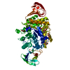 3vgdC 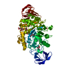 3vgeC  3vgfC 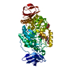 3vggC 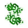 1eh9S S: Starting model for refinement C: citing same article ( |
|---|---|
| Similar structure data |


-Links
 PDBj
PDBj


- Assembly
| Deposited unit | 
| ||||||||
|---|---|---|---|---|---|---|---|---|---|
| 1 |
| ||||||||
| 2 | 
| ||||||||
| Unit cell |
|
-Components
| #1: Protein | Mass: 64740.754 Da / Num. of mol.: 1 / Mutation: E283Q Source method: isolated from a genetically manipulated source Source: (gene. exp.) Sulfolobus solfataricus (archaea) / Gene: treZ / Plasmid: PGUSS2 / Production host:  Pichia jadinii (fungus) Pichia jadinii (fungus)References: UniProt: Q55088, 4-alpha-D-{(1->4)-alpha-D-glucano}trehalose trehalohydrolase | ||||
|---|---|---|---|---|---|
| #2: Polysaccharide | alpha-D-glucopyranose-(1-1)-alpha-D-glucopyranose-(1-4)-alpha-D-glucopyranose-(1-4)-alpha-D- ...alpha-D-glucopyranose-(1-1)-alpha-D-glucopyranose-(1-4)-alpha-D-glucopyranose-(1-4)-alpha-D-glucopyranose-(1-4)-alpha-D-glucopyranose Source method: isolated from a genetically manipulated source | ||||
| #3: Chemical | ChemComp-FLC /   Mass: 189.100 Da / Num. of mol.: 1 / Source method: obtained synthetically / Formula: C6H5O7 Mass: 189.100 Da / Num. of mol.: 1 / Source method: obtained synthetically / Formula: C6H5O7 | ||||
| #4: Chemical |   Mass: 92.094 Da / Num. of mol.: 3 / Source method: obtained synthetically / Formula: C3H8O3 Mass: 92.094 Da / Num. of mol.: 3 / Source method: obtained synthetically / Formula: C3H8O3#5: Water | ChemComp-HOH / |  Mass: 18.015 Da / Num. of mol.: 163 / Source method: isolated from a natural source / Formula: H2O Mass: 18.015 Da / Num. of mol.: 163 / Source method: isolated from a natural source / Formula: H2OHas protein modification | Y | |
-Experimental details
-Experiment
| Experiment | Method: X-RAY DIFFRACTION / Number of used crystals: 1 |
|---|
- Sample preparation
| Crystal | Density Matthews: 3.91 Å3/Da / Density % sol: 68.57 % |
|---|---|
| Crystal grow | Temperature: 277 K / Method: vapor diffusion, hanging drop / pH: 7.5 Details: 1.1M sodium citrate, 0.1M HEPES, 5mM MTT, pH 7.5, VAPOR DIFFUSION, HANGING DROP, temperature 277K |
-Data collection
| Diffraction | Mean temperature: 100 K |
|---|---|
| Diffraction source | Source: SYNCHROTRON / Site: SPring-8  / Beamline: BL41XU / Wavelength: 1 Å / Beamline: BL41XU / Wavelength: 1 Å |
| Detector | Type: MAR CCD 165 mm / Detector: CCD / Date: Jan 24, 2001 / Details: mirrors |
| Radiation | Monochromator: Si 111 / Protocol: SINGLE WAVELENGTH / Monochromatic (M) / Laue (L): M / Scattering type: x-ray |
| Radiation wavelength | Wavelength: 1 Å / Relative weight: 1 |
| Reflection | Resolution: 2.6→55.3 Å / Num. obs: 31666 / % possible obs: 97.9 % / Redundancy: 7.3 % / Rmerge(I) obs: 0.099 / Net I/σ(I): 13.2 |
| Reflection shell | Resolution: 2.6→2.69 Å / Redundancy: 4.7 % / Rmerge(I) obs: 0.296 / Mean I/σ(I) obs: 1.7 / % possible all: 81.7 |
- Processing
| Software |
| |||||||||||||||||||||||||||||||||||||||||||||||||||||||||||||||||
|---|---|---|---|---|---|---|---|---|---|---|---|---|---|---|---|---|---|---|---|---|---|---|---|---|---|---|---|---|---|---|---|---|---|---|---|---|---|---|---|---|---|---|---|---|---|---|---|---|---|---|---|---|---|---|---|---|---|---|---|---|---|---|---|---|---|---|
| Refinement | Method to determine structure: MOLECULAR REPLACEMENT Starting model: PDB ENTRY 1EH9 Resolution: 2.6→55.27 Å / Cor.coef. Fo:Fc: 0.944 / Cor.coef. Fo:Fc free: 0.905 / WRfactor Rfree: 0.212 / WRfactor Rwork: 0.168 / Occupancy max: 1 / Occupancy min: 1 / FOM work R set: 0.8372 / SU B: 20.066 / SU ML: 0.192 / SU R Cruickshank DPI: 0.3451 / SU Rfree: 0.2605 / Cross valid method: THROUGHOUT / σ(F): 0 / ESU R Free: 0.26 / Stereochemistry target values: MAXIMUM LIKELIHOOD Details: HYDROGENS HAVE BEEN ADDED IN THE RIDING POSITIONS U VALUES
| |||||||||||||||||||||||||||||||||||||||||||||||||||||||||||||||||
| Solvent computation | Ion probe radii: 0.8 Å / Shrinkage radii: 0.8 Å / VDW probe radii: 1.4 Å / Solvent model: MASK | |||||||||||||||||||||||||||||||||||||||||||||||||||||||||||||||||
| Displacement parameters | Biso max: 108.31 Å2 / Biso mean: 45.7887 Å2 / Biso min: 16.16 Å2
| |||||||||||||||||||||||||||||||||||||||||||||||||||||||||||||||||
| Refinement step | Cycle: LAST / Resolution: 2.6→55.27 Å
| |||||||||||||||||||||||||||||||||||||||||||||||||||||||||||||||||
| Refine LS restraints |
| |||||||||||||||||||||||||||||||||||||||||||||||||||||||||||||||||
| LS refinement shell | Resolution: 2.601→2.669 Å / Total num. of bins used: 20
| |||||||||||||||||||||||||||||||||||||||||||||||||||||||||||||||||
| Refinement TLS params. | Method: refined / Origin x: 11.7367 Å / Origin y: 33.686 Å / Origin z: 22.5008 Å
|
