- PDB-3va7: Crystal structure of the Kluyveromyces lactis Urea Carboxylase -
+
Open data
ID or keywords:
Loading...
-
Basic information
Entry
Database: PDB / ID: 3va7
Title
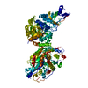
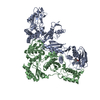
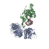
Crystal structure of the Kluyveromyces lactis Urea Carboxylase
Components
KLLA0E08119p
Keywords
LIGASE / Carboxylase
Function / homology
Function and homology information
urea carboxylase / urea carboxylase activity / allophanate hydrolase / allophanate hydrolase activity / small molecule metabolic process / ATP binding / metal ion binding Similarity search - Function
Urea carboxylase / Allophanate hydrolase / : / Allophanate hydrolase C-terminal domain / Carboxyltransferase domain, subdomain A and B / Carboxyltransferase domain, subdomain A and B / Allophanate hydrolase subunit 2 / Carboxyltransferase domain, subdomain C and D / Carboxyltransferase domain, subdomain C and D / Allophanate hydrolase subunit 1 ...Urea carboxylase / Allophanate hydrolase / : / Allophanate hydrolase C-terminal domain / Carboxyltransferase domain, subdomain A and B / Carboxyltransferase domain, subdomain A and B / Allophanate hydrolase subunit 2 / Carboxyltransferase domain, subdomain C and D / Carboxyltransferase domain, subdomain C and D / Allophanate hydrolase subunit 1 / Gyrase A; domain 2 - #40 / Amidase signature domain / Amidase signature (AS) superfamily / Amidase / : / Rossmann fold - #20 / Biotin-binding site / Biotin-requiring enzymes attachment site. / Cyclophilin-like / Cyclophilin / Biotin carboxylase-like, N-terminal domain / Biotin carboxylase, C-terminal / Biotin carboxylation domain / Biotin carboxylase, N-terminal domain / Biotin carboxylase C-terminal domain / Biotin carboxylation domain profile. / Biotin carboxylase C-terminal domain / Carbamoyl-phosphate synthase subdomain signature 1. / RNA polymerase II/Efflux pump adaptor protein, barrel-sandwich hybrid domain / Carbamoyl-phosphate synthetase large subunit-like, ATP-binding domain / Carbamoyl-phosphate synthase L chain, ATP binding domain / Biotin-requiring enzyme / Rudiment single hybrid motif / Biotinyl/lipoyl domain profile. / Biotin/lipoyl attachment / Single hybrid motif / ATP-grasp fold, B domain / Pre-ATP-grasp domain superfamily / Gyrase A; domain 2 / Cyclophilin-like domain superfamily / ATP-grasp fold / ATP-grasp fold profile. / D-amino Acid Aminotransferase; Chain A, domain 1 / OB fold (Dihydrolipoamide Acetyltransferase, E2P) / Carbamoyl-phosphate synthase subdomain signature 2. / Beta Barrel / Rossmann fold / 2-Layer Sandwich / 3-Layer(aba) Sandwich / Mainly Beta / Alpha Beta Similarity search - Domain/homology
Method to determine structure: SAD / Resolution: 2.6→50 Å / Cor.coef. Fo:Fc: 0.943 / Cor.coef. Fo:Fc free: 0.898 / SU B: 8.968 / SU ML: 0.195 / Cross valid method: THROUGHOUT / ESU R: 0.392 / ESU R Free: 0.286 / Stereochemistry target values: MAXIMUM LIKELIHOOD / Details: HYDROGENS HAVE BEEN ADDED IN THE RIDING POSITIONS
Rfactor
Num. reflection
% reflection
Selection details
Rfree
0.25524
2790
5.1 %
RANDOM
Rwork
0.18738
-
-
-
obs
0.19081
51949
99.12 %
-
Solvent computation
Ion probe radii: 0.8 Å / Shrinkage radii: 0.8 Å / VDW probe radii: 1.4 Å / Solvent model: MASK
Displacement parameters
Biso mean: 40.426 Å2
Baniso -1
Baniso -2
Baniso -3
1-
1.48 Å2
0 Å2
0 Å2
2-
-
1.48 Å2
0 Å2
3-
-
-
-2.96 Å2
Refinement step
Cycle: LAST / Resolution: 2.6→50 Å
Protein
Nucleic acid
Ligand
Solvent
Total
Num. atoms
8829
0
62
377
9268
Refine LS restraints
Refine-ID
Type
Dev ideal
Dev ideal target
Number
X-RAY DIFFRACTION
r_bond_refined_d
0.02
0.022
9072
X-RAY DIFFRACTION
r_bond_other_d
X-RAY DIFFRACTION
r_angle_refined_deg
1.922
1.97
12284
X-RAY DIFFRACTION
r_angle_other_deg
X-RAY DIFFRACTION
r_dihedral_angle_1_deg
7.183
5
1127
X-RAY DIFFRACTION
r_dihedral_angle_2_deg
38.311
24.512
410
X-RAY DIFFRACTION
r_dihedral_angle_3_deg
20.598
15
1546
X-RAY DIFFRACTION
r_dihedral_angle_4_deg
19.075
15
53
X-RAY DIFFRACTION
r_chiral_restr
0.13
0.2
1353
X-RAY DIFFRACTION
r_gen_planes_refined
0.008
0.021
6872
X-RAY DIFFRACTION
r_gen_planes_other
X-RAY DIFFRACTION
r_nbd_refined
X-RAY DIFFRACTION
r_nbd_other
X-RAY DIFFRACTION
r_nbtor_refined
X-RAY DIFFRACTION
r_nbtor_other
X-RAY DIFFRACTION
r_xyhbond_nbd_refined
X-RAY DIFFRACTION
r_xyhbond_nbd_other
X-RAY DIFFRACTION
r_metal_ion_refined
X-RAY DIFFRACTION
r_metal_ion_other
X-RAY DIFFRACTION
r_symmetry_vdw_refined
X-RAY DIFFRACTION
r_symmetry_vdw_other
X-RAY DIFFRACTION
r_symmetry_hbond_refined
X-RAY DIFFRACTION
r_symmetry_hbond_other
X-RAY DIFFRACTION
r_symmetry_metal_ion_refined
X-RAY DIFFRACTION
r_symmetry_metal_ion_other
X-RAY DIFFRACTION
r_mcbond_it
0.889
1.5
5622
X-RAY DIFFRACTION
r_mcbond_other
X-RAY DIFFRACTION
r_mcangle_it
1.704
2
9078
X-RAY DIFFRACTION
r_scbond_it
2.708
3
3450
X-RAY DIFFRACTION
r_scangle_it
4.373
4.5
3206
X-RAY DIFFRACTION
r_rigid_bond_restr
X-RAY DIFFRACTION
r_sphericity_free
X-RAY DIFFRACTION
r_sphericity_bonded
LS refinement shell
Resolution: 2.6→2.667 Å / Total num. of bins used: 20
Rfactor
Num. reflection
% reflection
Rfree
0.335
211
-
Rwork
0.264
3756
-
obs
-
-
99.13 %
+
About Yorodumi
-
News
-
Feb 9, 2022. New format data for meta-information of EMDB entries
New format data for meta-information of EMDB entries
Version 3 of the EMDB header file is now the official format.
The previous official version 1.9 will be removed from the archive.
In the structure databanks used in Yorodumi, some data are registered as the other names, "COVID-19 virus" and "2019-nCoV". Here are the details of the virus and the list of structure data.
Jan 31, 2019. EMDB accession codes are about to change! (news from PDBe EMDB page)
EMDB accession codes are about to change! (news from PDBe EMDB page)
The allocation of 4 digits for EMDB accession codes will soon come to an end. Whilst these codes will remain in use, new EMDB accession codes will include an additional digit and will expand incrementally as the available range of codes is exhausted. The current 4-digit format prefixed with “EMD-” (i.e. EMD-XXXX) will advance to a 5-digit format (i.e. EMD-XXXXX), and so on. It is currently estimated that the 4-digit codes will be depleted around Spring 2019, at which point the 5-digit format will come into force.
The EM Navigator/Yorodumi systems omit the EMD- prefix.
Related info.:Q: What is EMD? / ID/Accession-code notation in Yorodumi/EM Navigator
Yorodumi is a browser for structure data from EMDB, PDB, SASBDB, etc.
This page is also the successor to EM Navigator detail page, and also detail information page/front-end page for Omokage search.
The word "yorodu" (or yorozu) is an old Japanese word meaning "ten thousand". "mi" (miru) is to see.
Related info.:EMDB / PDB / SASBDB / Comparison of 3 databanks / Yorodumi Search / Aug 31, 2016. New EM Navigator & Yorodumi / Yorodumi Papers / Jmol/JSmol / Function and homology information / Changes in new EM Navigator and Yorodumi
 Movie
Movie Controller


 Open data
 Basic information
 Components
 Keywords
 Function and homology information
 Kluyveromyces lactis (yeast)
 X-RAY DIFFRACTION /
  Authors
 Citation
 Structure visualization
 Downloads & links
 Other downloads


 PDBj
PDBj


 Assembly


 Mass: 228.311 Da / Num. of mol.: 1 / Source method: obtained synthetically / Formula: C10H16N2O2S
 Mass: 60.055 Da / Num. of mol.: 1 / Source method: obtained synthetically / Formula: CH4N2O
 Mass: 92.094 Da / Num. of mol.: 7 / Source method: obtained synthetically / Formula: C3H8O3
 Mass: 22.990 Da / Num. of mol.: 1 / Source method: obtained synthetically / Formula: Na
 Sample preparation
 / Beamline: BL17U / Wavelength: 0.9796 Å
 Processing