SUMOylation of DNA damage response and repair proteins / G2/M DNA damage checkpoint / positive regulation of anoikis / Nonhomologous End-Joining (NHEJ) / mitotic DNA damage checkpoint signaling / Processing of DNA double-strand break ends / Recruitment and ATM-mediated phosphorylation of repair and signaling proteins at DNA double strand breaks / cellular response to bisphenol A / regulation of autophagosome assembly / DNA replication checkpoint signaling ...SUMOylation of DNA damage response and repair proteins / G2/M DNA damage checkpoint / positive regulation of anoikis / Nonhomologous End-Joining (NHEJ) / mitotic DNA damage checkpoint signaling / Processing of DNA double-strand break ends / Recruitment and ATM-mediated phosphorylation of repair and signaling proteins at DNA double strand breaks / cellular response to bisphenol A / regulation of autophagosome assembly / DNA replication checkpoint signaling / chromatin-protein adaptor activity / protein localization to site of double-strand break / mitotic intra-S DNA damage checkpoint signaling / response to glycoside / thymocyte apoptotic process / cellular response to stress / regulation of protein catabolic process / histone reader activity / negative regulation of DNA damage checkpoint / replicative senescence / signal transduction in response to DNA damage / intrinsic apoptotic signaling pathway in response to DNA damage by p53 class mediator / mitotic spindle assembly / Chk1/Chk2(Cds1) mediated inactivation of Cyclin B:Cdk1 complex / DNA damage checkpoint signaling / regulation of signal transduction by p53 class mediator / Ubiquitin-Mediated Degradation of Phosphorylated Cdc25A / DNA damage response, signal transduction by p53 class mediator / Stabilization of p53 / protein catabolic process / cellular response to gamma radiation / PML body / G2/M DNA damage checkpoint / Regulation of TP53 Activity through Methylation / G2/M transition of mitotic cell cycle / cellular response to xenobiotic stimulus / intrinsic apoptotic signaling pathway in response to DNA damage / Regulation of TP53 Degradation / double-strand break repair / Recruitment and ATM-mediated phosphorylation of repair and signaling proteins at DNA double strand breaks / chromosome / site of double-strand break / protein autophosphorylation / Regulation of TP53 Activity through Phosphorylation / protein phosphorylation / non-specific serine/threonine protein kinase / protein stabilization / cell division / protein serine kinase activity / DNA repair / protein serine/threonine kinase activity / DNA damage response / ubiquitin protein ligase binding / regulation of DNA-templated transcription / protein kinase binding / positive regulation of DNA-templated transcription / Golgi apparatus / protein homodimerization activity / nucleoplasm / ATP binding / metal ion binding / identical protein binding / nucleus / cytoplasm Similarity search - Function
Resolution: 1.54→1.6 Å / Redundancy: 4.5 % / Rmerge(I) obs: 0.508 / Mean I/σ(I) obs: 2.2 / Num. unique all: 3039 / Rsym value: 0.508 / % possible all: 99.3
-
Processing
Software
Name
Version
Classification
HKL-2000
datacollection
SHELXS
phasing
REFMAC
5.6.0117
refinement
HKL-2000
datareduction
HKL-2000
datascaling
Refinement
Method to determine structure: SAD / Resolution: 1.54→50 Å / Cor.coef. Fo:Fc: 0.969 / Cor.coef. Fo:Fc free: 0.958 / SU B: 3.104 / SU ML: 0.051 / Cross valid method: THROUGHOUT / ESU R: 0.102 / ESU R Free: 0.083 / Stereochemistry target values: MAXIMUM LIKELIHOOD / Details: HYDROGENS HAVE BEEN USED IF PRESENT IN THE INPUT
Rfactor
Num. reflection
% reflection
Selection details
Rfree
0.20465
1546
5 %
RANDOM
Rwork
0.15925
-
-
-
all
0.1616
31042
-
-
obs
0.1616
29179
98.98 %
-
Solvent computation
Ion probe radii: 0.8 Å / Shrinkage radii: 0.8 Å / VDW probe radii: 1.2 Å / Solvent model: MASK
Displacement parameters
Biso mean: 23.648 Å2
Baniso -1
Baniso -2
Baniso -3
1-
-0.91 Å2
0 Å2
-0 Å2
2-
-
-0.01 Å2
0 Å2
3-
-
-
0.92 Å2
Refinement step
Cycle: LAST / Resolution: 1.54→50 Å
Protein
Nucleic acid
Ligand
Solvent
Total
Num. atoms
1782
0
0
153
1935
Refine LS restraints
Refine-ID
Type
Dev ideal
Dev ideal target
Number
X-RAY DIFFRACTION
r_bond_refined_d
0.022
0.019
1833
X-RAY DIFFRACTION
r_angle_refined_deg
2.293
1.987
2498
X-RAY DIFFRACTION
r_dihedral_angle_1_deg
6.665
5
224
X-RAY DIFFRACTION
r_dihedral_angle_2_deg
39.372
23.25
80
X-RAY DIFFRACTION
r_dihedral_angle_3_deg
16.827
15
291
X-RAY DIFFRACTION
r_dihedral_angle_4_deg
23.767
15
14
X-RAY DIFFRACTION
r_chiral_restr
0.171
0.2
272
X-RAY DIFFRACTION
r_gen_planes_refined
0.015
0.022
1414
X-RAY DIFFRACTION
r_rigid_bond_restr
7.855
3
1833
X-RAY DIFFRACTION
r_sphericity_free
20.219
5
62
X-RAY DIFFRACTION
r_sphericity_bonded
13.584
5
1873
LS refinement shell
Resolution: 1.54→1.58 Å / Total num. of bins used: 20
Rfactor
Num. reflection
% reflection
Rfree
0.261
109
-
Rwork
0.18
1951
-
obs
-
-
98.71 %
Refinement TLS params.
Method: refined / Refine-ID: X-RAY DIFFRACTION
ID
L11 (°2)
L12 (°2)
L13 (°2)
L22 (°2)
L23 (°2)
L33 (°2)
S11 (Å °)
S12 (Å °)
S13 (Å °)
S21 (Å °)
S22 (Å °)
S23 (Å °)
S31 (Å °)
S32 (Å °)
S33 (Å °)
T11 (Å2)
T12 (Å2)
T13 (Å2)
T22 (Å2)
T23 (Å2)
T33 (Å2)
Origin x (Å)
Origin y (Å)
Origin z (Å)
1
1.0035
0.0097
0.3494
0.0132
-0.0939
1.1976
-0.0627
-0.0259
0.0818
0.0007
-0.0157
-0.0041
0.0369
0.0101
0.0784
0.0307
-0.0124
-0.008
0.0315
-0.0016
0.0114
19.3651
33.6512
26.1557
2
0.9025
0.1643
0.0262
0.4103
0.0129
0.5273
0.0005
-0.024
0.0076
-0.0565
-0.0063
-0.0373
-0.0255
-0.0169
0.0058
0.0119
0.0054
0.0077
0.0099
0.0017
0.0273
17.3051
36.3626
1.0532
Refinement TLS group
ID
Refine-ID
Refine TLS-ID
Auth asym-ID
Auth seq-ID
1
X-RAY DIFFRACTION
1
A
27 - 137
2
X-RAY DIFFRACTION
1
A
201 - 263
3
X-RAY DIFFRACTION
2
B
28 - 135
4
X-RAY DIFFRACTION
2
B
201 - 279
+
About Yorodumi
-
News
-
Feb 9, 2022. New format data for meta-information of EMDB entries
New format data for meta-information of EMDB entries
Version 3 of the EMDB header file is now the official format.
The previous official version 1.9 will be removed from the archive.
In the structure databanks used in Yorodumi, some data are registered as the other names, "COVID-19 virus" and "2019-nCoV". Here are the details of the virus and the list of structure data.
Jan 31, 2019. EMDB accession codes are about to change! (news from PDBe EMDB page)
EMDB accession codes are about to change! (news from PDBe EMDB page)
The allocation of 4 digits for EMDB accession codes will soon come to an end. Whilst these codes will remain in use, new EMDB accession codes will include an additional digit and will expand incrementally as the available range of codes is exhausted. The current 4-digit format prefixed with “EMD-” (i.e. EMD-XXXX) will advance to a 5-digit format (i.e. EMD-XXXXX), and so on. It is currently estimated that the 4-digit codes will be depleted around Spring 2019, at which point the 5-digit format will come into force.
The EM Navigator/Yorodumi systems omit the EMD- prefix.
Related info.:Q: What is EMD? / ID/Accession-code notation in Yorodumi/EM Navigator
Yorodumi is a browser for structure data from EMDB, PDB, SASBDB, etc.
This page is also the successor to EM Navigator detail page, and also detail information page/front-end page for Omokage search.
The word "yorodu" (or yorozu) is an old Japanese word meaning "ten thousand". "mi" (miru) is to see.
Related info.:EMDB / PDB / SASBDB / Comparison of 3 databanks / Yorodumi Search / Aug 31, 2016. New EM Navigator & Yorodumi / Yorodumi Papers / Jmol/JSmol / Function and homology information / Changes in new EM Navigator and Yorodumi
 Movie
Movie Controller

 Yorodumi
 Open data
 Basic information
 Components
 Keywords
 Function and homology information
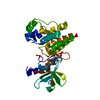
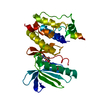
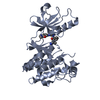
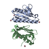
 Homo sapiens (human)
 X-RAY DIFFRACTION /
  Authors
 Citation
 Structure visualization
 Downloads & links
 Other downloads


 PDBj
PDBj


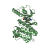
 Assembly
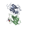

 Mass: 18.015 Da / Num. of mol.: 153 / Source method: isolated from a natural source / Formula: H2O
 Sample preparation
 / Beamline: BL13B1 / Wavelength: 0.9762 Å
 Processing