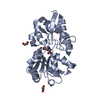
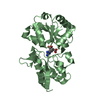
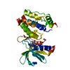
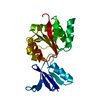
SUMOylation of DNA damage response and repair proteins / G2/M DNA damage checkpoint / Nonhomologous End-Joining (NHEJ) / Processing of DNA double-strand break ends / Recruitment and ATM-mediated phosphorylation of repair and signaling proteins at DNA double strand breaks / DNA replication checkpoint signaling / protein localization to site of double-strand break / protein K6-linked ubiquitination / chromatin-protein adaptor activity / histone reader activity ...SUMOylation of DNA damage response and repair proteins / G2/M DNA damage checkpoint / Nonhomologous End-Joining (NHEJ) / Processing of DNA double-strand break ends / Recruitment and ATM-mediated phosphorylation of repair and signaling proteins at DNA double strand breaks / DNA replication checkpoint signaling / protein localization to site of double-strand break / protein K6-linked ubiquitination / chromatin-protein adaptor activity / histone reader activity / positive regulation of double-strand break repair via homologous recombination / chromosome / site of double-strand break / nuclear body / focal adhesion / DNA repair / DNA damage response / nucleoplasm / nucleus Similarity search - Function
Resolution: 1.75→1.81 Å / Redundancy: 5.5 % / Rmerge(I) obs: 0.498 / Mean I/σ(I) obs: 2.9 / Num. unique all: 2386 / Rsym value: 0.498 / % possible all: 96
-
Processing
Software
Name
Version
Classification
HKL-2000
datacollection
SHELXS
phasing
REFMAC
5.6.0117
refinement
HKL-2000
datareduction
HKL-2000
datascaling
Refinement
Method to determine structure: SAD / Resolution: 1.74→50 Å / Cor.coef. Fo:Fc: 0.962 / Cor.coef. Fo:Fc free: 0.929 / SU B: 7.31 / SU ML: 0.102 / Cross valid method: THROUGHOUT / ESU R: 0.172 / ESU R Free: 0.136 / Stereochemistry target values: MAXIMUM LIKELIHOOD / Details: HYDROGENS HAVE BEEN USED IF PRESENT IN THE INPUT
Rfactor
Num. reflection
% reflection
Selection details
Rfree
0.27176
1265
5.1 %
RANDOM
Rwork
0.18861
-
-
-
all
0.19273
25501
-
-
obs
0.19273
23441
96.88 %
-
Solvent computation
Ion probe radii: 0.8 Å / Shrinkage radii: 0.8 Å / VDW probe radii: 1.2 Å / Solvent model: MASK
Displacement parameters
Biso mean: 28.364 Å2
Baniso -1
Baniso -2
Baniso -3
1-
-0.27 Å2
-0 Å2
0 Å2
2-
-
2.38 Å2
-0 Å2
3-
-
-
-2.11 Å2
Refinement step
Cycle: LAST / Resolution: 1.74→50 Å
Protein
Nucleic acid
Ligand
Solvent
Total
Num. atoms
1675
0
10
146
1831
Refine LS restraints
Refine-ID
Type
Dev ideal
Dev ideal target
Number
X-RAY DIFFRACTION
r_bond_refined_d
0.019
0.019
1723
X-RAY DIFFRACTION
r_angle_refined_deg
2.055
1.982
2346
X-RAY DIFFRACTION
r_dihedral_angle_1_deg
6.132
5
212
X-RAY DIFFRACTION
r_dihedral_angle_2_deg
37.95
23.158
76
X-RAY DIFFRACTION
r_dihedral_angle_3_deg
17.103
15
276
X-RAY DIFFRACTION
r_dihedral_angle_4_deg
17.227
15
14
X-RAY DIFFRACTION
r_chiral_restr
0.138
0.2
255
X-RAY DIFFRACTION
r_gen_planes_refined
0.011
0.022
1336
X-RAY DIFFRACTION
r_rigid_bond_restr
7.105
3
1723
X-RAY DIFFRACTION
r_sphericity_free
23.092
5
65
X-RAY DIFFRACTION
r_sphericity_bonded
13.596
5
1756
LS refinement shell
Resolution: 1.745→1.79 Å / Total num. of bins used: 20
Rfactor
Num. reflection
% reflection
Rfree
0.42
77
-
Rwork
0.275
1411
-
obs
-
1616
87.32 %
Refinement TLS params.
Method: refined / Refine-ID: X-RAY DIFFRACTION
ID
L11 (°2)
L12 (°2)
L13 (°2)
L22 (°2)
L23 (°2)
L33 (°2)
S11 (Å °)
S12 (Å °)
S13 (Å °)
S21 (Å °)
S22 (Å °)
S23 (Å °)
S31 (Å °)
S32 (Å °)
S33 (Å °)
T11 (Å2)
T12 (Å2)
T13 (Å2)
T22 (Å2)
T23 (Å2)
T33 (Å2)
Origin x (Å)
Origin y (Å)
Origin z (Å)
1
1.6606
0.2605
-0.7699
1.7186
0.17
1.7689
0.1204
-0.1987
0.086
0.0812
-0.044
-0.0165
-0.1033
0.0948
-0.0765
0.0231
-0.0144
0.02
0.0722
-0.0053
0.0253
1.4479
6.1664
24.1078
2
2.0842
0.1394
0.0473
2.0534
-0.4162
2.8613
0.044
0.0393
-0.2525
-0.3692
-0.0636
0.0871
0.2567
-0.1249
0.0195
0.106
0.0093
-0.0121
0.031
-0.0063
0.0401
-13.0547
-0.518
5.6736
Refinement TLS group
ID
Refine-ID
Refine TLS-ID
Auth asym-ID
Auth seq-ID
1
X-RAY DIFFRACTION
1
A
29 - 135
2
X-RAY DIFFRACTION
1
A
201 - 383
3
X-RAY DIFFRACTION
2
B
28 - 134
4
X-RAY DIFFRACTION
2
B
201 - 363
+
About Yorodumi
-
News
-
Feb 9, 2022. New format data for meta-information of EMDB entries
New format data for meta-information of EMDB entries
Version 3 of the EMDB header file is now the official format.
The previous official version 1.9 will be removed from the archive.
In the structure databanks used in Yorodumi, some data are registered as the other names, "COVID-19 virus" and "2019-nCoV". Here are the details of the virus and the list of structure data.
Jan 31, 2019. EMDB accession codes are about to change! (news from PDBe EMDB page)
EMDB accession codes are about to change! (news from PDBe EMDB page)
The allocation of 4 digits for EMDB accession codes will soon come to an end. Whilst these codes will remain in use, new EMDB accession codes will include an additional digit and will expand incrementally as the available range of codes is exhausted. The current 4-digit format prefixed with “EMD-” (i.e. EMD-XXXX) will advance to a 5-digit format (i.e. EMD-XXXXX), and so on. It is currently estimated that the 4-digit codes will be depleted around Spring 2019, at which point the 5-digit format will come into force.
The EM Navigator/Yorodumi systems omit the EMD- prefix.
Related info.:Q: What is EMD? / ID/Accession-code notation in Yorodumi/EM Navigator
Yorodumi is a browser for structure data from EMDB, PDB, SASBDB, etc.
This page is also the successor to EM Navigator detail page, and also detail information page/front-end page for Omokage search.
The word "yorodu" (or yorozu) is an old Japanese word meaning "ten thousand". "mi" (miru) is to see.
Related info.:EMDB / PDB / SASBDB / Comparison of 3 databanks / Yorodumi Search / Aug 31, 2016. New EM Navigator & Yorodumi / Yorodumi Papers / Jmol/JSmol / Function and homology information / Changes in new EM Navigator and Yorodumi
 Movie
Movie Controller


 Open data
 Basic information
 Components
 Keywords
 Function and homology information
 X-RAY DIFFRACTION /
  Authors
 Citation
 Structure visualization
 Downloads & links
 Other downloads


 PDBj
PDBj


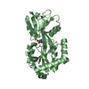
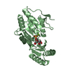
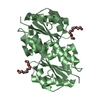
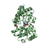
 Assembly


 Mass: 96.063 Da / Num. of mol.: 2 / Source method: obtained synthetically / Formula: SO4
 Mass: 18.015 Da / Num. of mol.: 146 / Source method: isolated from a natural source / Formula: H2O
 Sample preparation
 / Beamline: BL12B2 / Wavelength: 1 Å
 Processing