| 登録情報 | データベース: PDB / ID: 3v4q
|
|---|
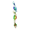
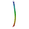
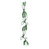
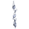
| タイトル | Structure of R335W mutant of human Lamin |
|---|
 要素 要素 | Prelamin-A/C |
|---|
 キーワード キーワード | STRUCTURAL PROTEIN |
|---|
| 機能・相同性 |  機能・相同性情報 機能・相同性情報
structural constituent of nuclear lamina / negative regulation of mesenchymal cell proliferation / establishment or maintenance of microtubule cytoskeleton polarity / Breakdown of the nuclear lamina / DNA double-strand break attachment to nuclear envelope / ventricular cardiac muscle cell development / Depolymerization of the Nuclear Lamina / nuclear envelope organization / Nuclear Envelope Breakdown / nuclear pore localization ...structural constituent of nuclear lamina / negative regulation of mesenchymal cell proliferation / establishment or maintenance of microtubule cytoskeleton polarity / Breakdown of the nuclear lamina / DNA double-strand break attachment to nuclear envelope / ventricular cardiac muscle cell development / Depolymerization of the Nuclear Lamina / nuclear envelope organization / Nuclear Envelope Breakdown / nuclear pore localization / lamin filament / protein localization to nuclear envelope / XBP1(S) activates chaperone genes / nuclear lamina / Initiation of Nuclear Envelope (NE) Reformation / regulation of protein localization to nucleus / intermediate filament / regulation of telomere maintenance / negative regulation of cardiac muscle hypertrophy in response to stress / nuclear migration / muscle organ development / Deregulated CDK5 triggers multiple neurodegenerative pathways in Alzheimer's disease models / negative regulation of release of cytochrome c from mitochondria / protein localization to nucleus / regulation of cell migration / Meiotic synapsis / negative regulation of extrinsic apoptotic signaling pathway / regulation of protein stability / structural constituent of cytoskeleton / nuclear matrix / protein import into nucleus / cellular senescence / Signaling by BRAF and RAF1 fusions / intracellular protein localization / nuclear envelope / heterochromatin formation / site of double-strand break / nuclear membrane / cellular response to hypoxia / nuclear speck / negative regulation of cell population proliferation / positive regulation of gene expression / perinuclear region of cytoplasm / structural molecule activity / nucleoplasm / identical protein binding / nucleus / cytosol類似検索 - 分子機能 Lamin tail domain superfamily / Lamin tail domain / Lamin Tail Domain / Lamin-tail (LTD) domain profile. / Intermediate filament protein, conserved site / Intermediate filament protein / Intermediate filament (IF) rod domain signature. / Intermediate filament, rod domain / Intermediate filament (IF) rod domain profile. / Intermediate filament protein ...Lamin tail domain superfamily / Lamin tail domain / Lamin Tail Domain / Lamin-tail (LTD) domain profile. / Intermediate filament protein, conserved site / Intermediate filament protein / Intermediate filament (IF) rod domain signature. / Intermediate filament, rod domain / Intermediate filament (IF) rod domain profile. / Intermediate filament protein / Single alpha-helices involved in coiled-coils or other helix-helix interfaces - #170 / Single alpha-helices involved in coiled-coils or other helix-helix interfaces / Up-down Bundle / Mainly Alpha類似検索 - ドメイン・相同性 |
|---|
| 生物種 |  Homo sapiens (ヒト) Homo sapiens (ヒト) |
|---|
| 手法 |  X線回折 / シンクロトロン / 分子置換 / 解像度: 3.06 Å X線回折 / シンクロトロン / 分子置換 / 解像度: 3.06 Å |
|---|
 データ登録者 データ登録者 | Bollati, M. / Bolognesi, M. |
|---|
 引用 引用 | ジャーナル: Biochem.Biophys.Res.Commun. / 年: 2012
タイトル: Structures of the lamin A/C R335W and E347K mutants: Implications for dilated cardiolaminopathies.
著者: Bollati, M. / Barbiroli, A. / Favalli, V. / Arbustini, E. / Charron, P. / Bolognesi, M. |
|---|
| 履歴 | | 登録 | 2011年12月15日 | 登録サイト: RCSB / 処理サイト: RCSB |
|---|
| 改定 1.0 | 2012年2月29日 | Provider: repository / タイプ: Initial release |
|---|
| 改定 1.1 | 2018年1月24日 | Group: Structure summary / カテゴリ: audit_author / Item: _audit_author.name |
|---|
| 改定 1.2 | 2018年6月20日 | Group: Data collection / カテゴリ: diffrn_radiation
Item: _diffrn_radiation.pdbx_diffrn_protocol / _diffrn_radiation.pdbx_scattering_type |
|---|
| 改定 1.3 | 2024年2月28日 | Group: Data collection / Database references
カテゴリ: chem_comp_atom / chem_comp_bond ...chem_comp_atom / chem_comp_bond / database_2 / struct_ref_seq_dif
Item: _database_2.pdbx_DOI / _database_2.pdbx_database_accession / _struct_ref_seq_dif.details |
|---|
|
|---|
 ムービー
ムービー コントローラー


 データを開く
 基本情報
基本情報 構造の表示
構造の表示 ダウンロードとリンク
 その他のダウンロード


 PDBj
PDBj


 集合体
集合体

 試料調製
試料調製 / ビームライン: ID23-1 / 波長: 0.9769 Å
 解析
解析