 Movie
Movie Controller

[English] 日本語
 Yorodumi
- PDB-3v0u: Crystal Structure of Perakine Reductase, Founder Member of a Nove... -
+ Open data

- Basic information
| Entry | Database: PDB / ID: 3v0u | ||||||
|---|---|---|---|---|---|---|---|
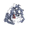
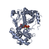
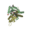
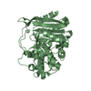
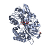
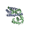
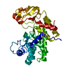
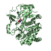
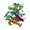
| Title | Crystal Structure of Perakine Reductase, Founder Member of a Novel AKR Subfamily with Unique Conformational Changes during NADPH Binding | ||||||
 Components Components | Perakine reductase | ||||||
 Keywords Keywords | OXIDOREDUCTASE / Perakine Reductase / AKR superfamily | ||||||
| Function / homology |  Function and homology information Function and homology informationperakine reductase / alkaloid metabolic process / oxidoreductase activity / cytoplasm Similarity search - Function | ||||||
| Biological species |  Rauvolfia serpentina (serpentwood) Rauvolfia serpentina (serpentwood) | ||||||
| Method |  X-RAY DIFFRACTION / SYNCHROTRON / MOLECULAR REPLACEMENT / Resolution: 2.203 Å X-RAY DIFFRACTION / SYNCHROTRON / MOLECULAR REPLACEMENT / Resolution: 2.203 Å | ||||||
 Authors Authors | Sun, L. / Chen, Y. / Rajendran, C. / Panjikar, S. / Mueller, U. / Wang, M. / Rosenthal, C. / Mindnich, R. / Penning, T.M. / Stoeckigt, J. | ||||||
 Citation Citation | Journal: J.Biol.Chem. / Year: 2012 Title: Crystal structure of perakine reductase, founding member of a novel aldo-keto reductase (AKR) subfamily that undergoes unique conformational changes during NADPH binding. Authors: Sun, L. / Chen, Y. / Rajendran, C. / Mueller, U. / Panjikar, S. / Wang, M. / Mindnich, R. / Rosenthal, C. / Penning, T.M. / Stockigt, J. | ||||||
| History |
|
- Structure visualization
| Structure viewer | Molecule: MolmilJmol/JSmol |
|---|
- Downloads & links
-Download
| PDBx/mmCIF format | 3v0u.cif.gz | 124.4 KB | Display | PDBx/mmCIF format |
|---|---|---|---|---|
| PDB format | pdb3v0u.ent.gz | 96.6 KB | Display | PDB format |
| PDBx/mmJSON format | 3v0u.json.gz | Tree view | PDBx/mmJSON format | |
| Others |  Other downloads Other downloads |
-Validation report
| Arichive directory | https://data.pdbj.org/pub/pdb/validation_reports/v0/3v0uftp://data.pdbj.org/pub/pdb/validation_reports/v0/3v0u | HTTPS FTP |
|---|
-Related structure data
| Related structure data |  3uyiC 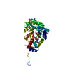 3v0sC 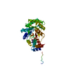 3v0tC  1pz0S C: citing same article ( S: Starting model for refinement |
|---|---|
| Similar structure data |

-Links
 PDBj
PDBj
- Assembly
| Deposited unit | 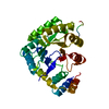
| ||||||||
|---|---|---|---|---|---|---|---|---|---|
| 1 |
| ||||||||
| 2 | 
| ||||||||
| Unit cell |
|
-Components
| #1: Protein | Mass: 37460.953 Da / Num. of mol.: 1 / Mutation: A213W Source method: isolated from a genetically manipulated source Source: (gene. exp.) Rauvolfia serpentina (serpentwood) / Gene: PR / Production host:  |
|---|---|
| #2: Water | ChemComp-HOH /  Mass: 18.015 Da / Num. of mol.: 69 / Source method: isolated from a natural source / Formula: H2O Mass: 18.015 Da / Num. of mol.: 69 / Source method: isolated from a natural source / Formula: H2O |
-Experimental details
-Experiment
| Experiment | Method: X-RAY DIFFRACTION / Number of used crystals: 1 |
|---|
- Sample preparation
| Crystal | Density Matthews: 2.6 Å3/Da / Density % sol: 52.65 % |
|---|
-Data collection
| Diffraction source | Source: SYNCHROTRON / Site: SLS  / Beamline: X06SA / Wavelength: 1 Å / Beamline: X06SA / Wavelength: 1 Å |
|---|---|
| Radiation | Protocol: SINGLE WAVELENGTH / Monochromatic (M) / Laue (L): M / Scattering type: x-ray |
| Radiation wavelength | Wavelength: 1 Å / Relative weight: 1 |
| Reflection | Resolution: 2.2→71.5 Å / Num. obs: 20584 / % possible obs: 95.9 % / Observed criterion σ(F): 2 / Observed criterion σ(I): 2 |
- Processing
| Software | Name: PHENIX / Version: (phenix.refine: 1.7.1_743) / Classification: refinement | |||||||||||||||||||||||||||||||||||||||||||||||||||||||||||||||||||||||||||
|---|---|---|---|---|---|---|---|---|---|---|---|---|---|---|---|---|---|---|---|---|---|---|---|---|---|---|---|---|---|---|---|---|---|---|---|---|---|---|---|---|---|---|---|---|---|---|---|---|---|---|---|---|---|---|---|---|---|---|---|---|---|---|---|---|---|---|---|---|---|---|---|---|---|---|---|---|
| Refinement | Method to determine structure: MOLECULAR REPLACEMENT Starting model: 1PZ0 Resolution: 2.203→40.634 Å / SU ML: 0.58 / σ(F): 2.01 / Phase error: 25.38 / Stereochemistry target values: ML
| |||||||||||||||||||||||||||||||||||||||||||||||||||||||||||||||||||||||||||
| Solvent computation | Shrinkage radii: 0.72 Å / VDW probe radii: 1 Å / Solvent model: FLAT BULK SOLVENT MODEL / Bsol: 54.957 Å2 / ksol: 0.338 e/Å3 | |||||||||||||||||||||||||||||||||||||||||||||||||||||||||||||||||||||||||||
| Displacement parameters |
| |||||||||||||||||||||||||||||||||||||||||||||||||||||||||||||||||||||||||||
| Refinement step | Cycle: LAST / Resolution: 2.203→40.634 Å
| |||||||||||||||||||||||||||||||||||||||||||||||||||||||||||||||||||||||||||
| Refine LS restraints |
| |||||||||||||||||||||||||||||||||||||||||||||||||||||||||||||||||||||||||||
| LS refinement shell | Refine-ID: X-RAY DIFFRACTION / Total num. of bins used: 7
| |||||||||||||||||||||||||||||||||||||||||||||||||||||||||||||||||||||||||||
| Refinement TLS params. | Method: refined / Refine-ID: X-RAY DIFFRACTION
| |||||||||||||||||||||||||||||||||||||||||||||||||||||||||||||||||||||||||||
| Refinement TLS group |
|
