 Movie
Movie Controller


+ Open data

- Basic information
| Entry | Database: PDB / ID: 3uu2 | ||||||
|---|---|---|---|---|---|---|---|
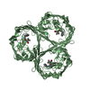
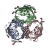
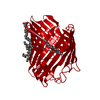
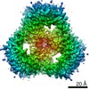
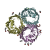
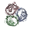
| Title | Salmonella typhi osmoporin(OmpC):an Outer Membrane Protein | ||||||
 Components Components | Outer membrane protein C | ||||||
 Keywords Keywords | MEMBRANE PROTEIN / Beta Barrel / Non specific porin / osmoporin / Outer Membrane | ||||||
| Function / homology |  Function and homology information Function and homology informationporin activity / pore complex / cell outer membrane / monoatomic ion transmembrane transport Similarity search - Function | ||||||
| Biological species |  Salmonella enterica subsp. enterica serovar Typhimurium (bacteria) Salmonella enterica subsp. enterica serovar Typhimurium (bacteria) | ||||||
| Method |  X-RAY DIFFRACTION / SYNCHROTRON / MOLECULAR REPLACEMENT / Resolution: 3.59 Å X-RAY DIFFRACTION / SYNCHROTRON / MOLECULAR REPLACEMENT / Resolution: 3.59 Å | ||||||
 Authors Authors | Prasanth, P. / Putcha, B.K. / Arockiasamy, A. / Krishnaswamy, S. | ||||||
 Citation Citation | Journal: To be Published Title: Crystal Structure of Outer membrane protein OmpC from Salmonella typhi. Authors: Prasanth, P. / Putcha, B.K. / Arockiasamy, A. / Krishnaswamy, S. | ||||||
| History |
|
- Structure visualization
| Structure viewer | Molecule: MolmilJmol/JSmol |
|---|
- Downloads & links
-Download
| PDBx/mmCIF format | 3uu2.cif.gz | 371.6 KB | Display | PDBx/mmCIF format |
|---|---|---|---|---|
| PDB format | pdb3uu2.ent.gz | 311.4 KB | Display | PDB format |
| PDBx/mmJSON format | 3uu2.json.gz | Tree view | PDBx/mmJSON format | |
| Others |  Other downloads Other downloads |
-Validation report
| Arichive directory | https://data.pdbj.org/pub/pdb/validation_reports/uu/3uu2ftp://data.pdbj.org/pub/pdb/validation_reports/uu/3uu2 | HTTPS FTP |
|---|
-Related structure data
| Related structure data |  3upgS S: Starting model for refinement |
|---|---|
| Similar structure data |


-Links
 PDBj
PDBj- Assembly
| Deposited unit | 
| ||||||||
|---|---|---|---|---|---|---|---|---|---|
| 1 |
| ||||||||
| Unit cell |
|
-Components
| #1: Protein | Mass: 39207.059 Da / Num. of mol.: 3 Source method: isolated from a genetically manipulated source Source: (gene. exp.) Salmonella enterica subsp. enterica serovar Typhimurium (bacteria)Strain: Ty21a / Gene: ompC, STM2267 / Plasmid: pET21b+ / Production host: |
|---|
-Experimental details
-Experiment
| Experiment | Method: X-RAY DIFFRACTION / Number of used crystals: 1 |
|---|
- Sample preparation
| Crystal | Density Matthews: 4.22 Å3/Da / Density % sol: 70.83 % |
|---|---|
| Crystal grow | Temperature: 298.15 K / Method: microbatch under oil / pH: 8.5 Details: 0.01M Nickel(II) chloride hexahydrate, 0.1M Tris, 20% PEG MME 2000, pH 8.5, Microbatch under oil, temperature 298.15K |
-Data collection
| Diffraction | Mean temperature: 77.03 K |
|---|---|
| Diffraction source | Source: SYNCHROTRON / Site: ESRF  / Beamline: BM14 / Wavelength: 1.0048 Å / Beamline: BM14 / Wavelength: 1.0048 Å |
| Detector | Type: MARMOSAIC 225 mm CCD / Detector: CCD / Date: May 4, 2010 |
| Radiation | Protocol: SINGLE WAVELENGTH / Monochromatic (M) / Laue (L): M / Scattering type: x-ray |
| Radiation wavelength | Wavelength: 1.0048 Å / Relative weight: 1 |
| Reflection | Resolution: 3.33→100.27 Å / Num. all: 28544 / Num. obs: 27261 / % possible obs: 95.7 % / Redundancy: 12.1 % / Biso Wilson estimate: 97.8 Å2 / Rmerge(I) obs: 0.11 / Net I/σ(I): 23 |
| Reflection shell | Resolution: 3.59→3.67 Å / Redundancy: 9.7 % / Rmerge(I) obs: 0.526 / Mean I/σ(I) obs: 2.95 / % possible all: 93.2 |
- Processing
| Software |
| ||||||||||||||||||||||||||||||||||||||||||||||||||||||||||||||||||||||||||||||||||||||||||||||||||||
|---|---|---|---|---|---|---|---|---|---|---|---|---|---|---|---|---|---|---|---|---|---|---|---|---|---|---|---|---|---|---|---|---|---|---|---|---|---|---|---|---|---|---|---|---|---|---|---|---|---|---|---|---|---|---|---|---|---|---|---|---|---|---|---|---|---|---|---|---|---|---|---|---|---|---|---|---|---|---|---|---|---|---|---|---|---|---|---|---|---|---|---|---|---|---|---|---|---|---|---|---|---|
| Refinement | Method to determine structure: MOLECULAR REPLACEMENT Starting model: 3UPG Resolution: 3.59→50 Å / Cor.coef. Fo:Fc: 0.876 / Cor.coef. Fo:Fc free: 0.848 / SU B: 130.247 / SU ML: 0.857 / Isotropic thermal model: Overall / Cross valid method: THROUGHOUT / ESU R Free: 0.748 / Stereochemistry target values: MAXIMUM LIKELIHOOD
| ||||||||||||||||||||||||||||||||||||||||||||||||||||||||||||||||||||||||||||||||||||||||||||||||||||
| Solvent computation | Ion probe radii: 0.8 Å / Shrinkage radii: 0.8 Å / VDW probe radii: 1.4 Å / Solvent model: MASK | ||||||||||||||||||||||||||||||||||||||||||||||||||||||||||||||||||||||||||||||||||||||||||||||||||||
| Displacement parameters | Biso mean: 225.198 Å2
| ||||||||||||||||||||||||||||||||||||||||||||||||||||||||||||||||||||||||||||||||||||||||||||||||||||
| Refinement step | Cycle: LAST / Resolution: 3.59→50 Å
| ||||||||||||||||||||||||||||||||||||||||||||||||||||||||||||||||||||||||||||||||||||||||||||||||||||
| Refine LS restraints |
| ||||||||||||||||||||||||||||||||||||||||||||||||||||||||||||||||||||||||||||||||||||||||||||||||||||
| LS refinement shell | Resolution: 3.59→3.683 Å / Total num. of bins used: 20
| ||||||||||||||||||||||||||||||||||||||||||||||||||||||||||||||||||||||||||||||||||||||||||||||||||||
| Refinement TLS params. | Method: refined / Refine-ID: X-RAY DIFFRACTION
| ||||||||||||||||||||||||||||||||||||||||||||||||||||||||||||||||||||||||||||||||||||||||||||||||||||
| Refinement TLS group |
|
