 Movie
Movie Controller

[English] 日本語
 Yorodumi
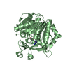
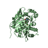
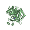
- PDB-3uq6: Adenosine kinase from Schistosoma mansoni in complex with adenosi... -
+ Open data

- Basic information
| Entry | Database: PDB / ID: 3uq6 | ||||||
|---|---|---|---|---|---|---|---|
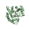
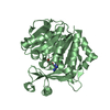
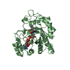
| Title | Adenosine kinase from Schistosoma mansoni in complex with adenosine and AMP | ||||||
 Components Components | Adenosine kinase, putative | ||||||
 Keywords Keywords | TRANSFERASE / Ribokinase | ||||||
| Function / homology |  Function and homology information Function and homology informationadenosine kinase / adenosine kinase activity / purine ribonucleoside salvage / nucleotide binding Similarity search - Function | ||||||
| Biological species |  | ||||||
| Method |  X-RAY DIFFRACTION / SYNCHROTRON / MOLECULAR REPLACEMENT / molecular replacement / Resolution: 2.3 Å X-RAY DIFFRACTION / SYNCHROTRON / MOLECULAR REPLACEMENT / molecular replacement / Resolution: 2.3 Å | ||||||
 Authors Authors | Romanello, L. / Cassago, A. / Bachega, F.R. / Garatt, R.C. / DeMarco, R. / Pereira, H.M. | ||||||
 Citation Citation | Journal: Acta Crystallogr.,Sect.D / Year: 2013 Title: Adenosine kinase from Schistosoma mansoni: structural basis for the differential incorporation of nucleoside analogues. Authors: Romanello, L. / Bachega, J.F. / Cassago, A. / Brandao-Neto, J. / Demarco, R. / Garratt, R.C. / Pereira, H.D. | ||||||
| History |
|
- Structure visualization
| Structure viewer | Molecule: MolmilJmol/JSmol |
|---|
- Downloads & links
-Download
| PDBx/mmCIF format | 3uq6.cif.gz | 278.6 KB | Display | PDBx/mmCIF format |
|---|---|---|---|---|
| PDB format | pdb3uq6.ent.gz | 224 KB | Display | PDB format |
| PDBx/mmJSON format | 3uq6.json.gz | Tree view | PDBx/mmJSON format | |
| Others |  Other downloads Other downloads |
-Validation report
| Arichive directory | https://data.pdbj.org/pub/pdb/validation_reports/uq/3uq6ftp://data.pdbj.org/pub/pdb/validation_reports/uq/3uq6 | HTTPS FTP |
|---|
-Related structure data
| Related structure data | 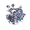 3uq9C 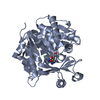 3vaqC 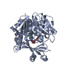 3vasC  4dc3C 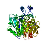 1bx4S C: citing same article ( S: Starting model for refinement |
|---|---|
| Similar structure data |


-Links
 PDBj
PDBj


- Assembly
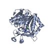
| Deposited unit | 
| ||||||||
|---|---|---|---|---|---|---|---|---|---|
| 1 | 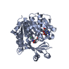
| ||||||||
| 2 | 
| ||||||||
| Unit cell |
|
-Components
| #1: Protein | Mass: 41609.617 Da / Num. of mol.: 2 Source method: isolated from a genetically manipulated source Details: 37oC / Source: (gene. exp.)  References: UniProt: C4PZB4, UniProt: G4V7G8*PLUS, adenosine kinase #2: Chemical |   Mass: 267.241 Da / Num. of mol.: 2 / Source method: obtained synthetically / Formula: C10H13N5O4 Mass: 267.241 Da / Num. of mol.: 2 / Source method: obtained synthetically / Formula: C10H13N5O4#3: Chemical |   Mass: 347.221 Da / Num. of mol.: 2 / Source method: obtained synthetically / Formula: C10H14N5O7P / Comment: AMP*YM Mass: 347.221 Da / Num. of mol.: 2 / Source method: obtained synthetically / Formula: C10H14N5O7P / Comment: AMP*YM#4: Chemical |   Mass: 35.453 Da / Num. of mol.: 2 / Source method: obtained synthetically / Formula: Cl Mass: 35.453 Da / Num. of mol.: 2 / Source method: obtained synthetically / Formula: Cl#5: Water | ChemComp-HOH / |  Mass: 18.015 Da / Num. of mol.: 184 / Source method: isolated from a natural source / Formula: H2O Mass: 18.015 Da / Num. of mol.: 184 / Source method: isolated from a natural source / Formula: H2O |
|---|
-Experimental details
-Experiment
| Experiment | Method: X-RAY DIFFRACTION / Number of used crystals: 1 |
|---|
- Sample preparation
| Crystal | Density Matthews: 2.55 Å3/Da / Density % sol: 51.73 % |
|---|---|
| Crystal grow | Temperature: 293 K Details: 100mM Bis-tris, 200mM LiSO4, 16-20% PEG 3350, VAPOR DIFFUSION, HANGING DROP, temperature 293K PH range: 6.1-6.7 |
-Data collection
| Diffraction | Mean temperature: 100 K |
|---|---|
| Diffraction source | Source: SYNCHROTRON / Site: LNLS  / Beamline: W01B-MX2 / Wavelength: 1.45 / Beamline: W01B-MX2 / Wavelength: 1.45 |
| Detector | Type: MARMOSAIC 225 mm CCD / Detector: CCD / Date: Sep 26, 2008 |
| Radiation | Monochromator: DCM SI(111) / Protocol: SINGLE WAVELENGTH / Monochromatic (M) / Laue (L): M / Scattering type: x-ray |
| Radiation wavelength | Wavelength: 1.45 Å / Relative weight: 1 |
| Reflection | Resolution: 2.3→180.53 Å / Num. obs: 35871 / % possible obs: 93.3 % / Observed criterion σ(I): 2 / Redundancy: 3.4 % / Rmerge(I) obs: 0.103 / Rsym value: 0.103 / Net I/σ(I): 10.7 |
| Reflection shell | Resolution: 2.3→2.42 Å / Redundancy: 3.4 % / Rmerge(I) obs: 0.552 / Mean I/σ(I) obs: 1.4 / Rsym value: 0.552 / % possible all: 88.6 |
-Phasing
| Phasing | Method: molecular replacement |
|---|
- Processing
| Software |
| ||||||||||||||||||||||||||||||||||||||||||||||||||||||||||||||||||||||||||||||||||||||||||||||||||
|---|---|---|---|---|---|---|---|---|---|---|---|---|---|---|---|---|---|---|---|---|---|---|---|---|---|---|---|---|---|---|---|---|---|---|---|---|---|---|---|---|---|---|---|---|---|---|---|---|---|---|---|---|---|---|---|---|---|---|---|---|---|---|---|---|---|---|---|---|---|---|---|---|---|---|---|---|---|---|---|---|---|---|---|---|---|---|---|---|---|---|---|---|---|---|---|---|---|---|---|
| Refinement | Method to determine structure: MOLECULAR REPLACEMENT Starting model: PDB ENTRY 1BX4 Resolution: 2.3→49.958 Å / Occupancy max: 1 / Occupancy min: 1 / SU ML: 0.25 / σ(F): 1.35 / Phase error: 25.57 / Stereochemistry target values: ML
| ||||||||||||||||||||||||||||||||||||||||||||||||||||||||||||||||||||||||||||||||||||||||||||||||||
| Solvent computation | Shrinkage radii: 0.9 Å / VDW probe radii: 1.11 Å / Solvent model: FLAT BULK SOLVENT MODEL | ||||||||||||||||||||||||||||||||||||||||||||||||||||||||||||||||||||||||||||||||||||||||||||||||||
| Displacement parameters | Biso mean: 43.24 Å2
| ||||||||||||||||||||||||||||||||||||||||||||||||||||||||||||||||||||||||||||||||||||||||||||||||||
| Refinement step | Cycle: LAST / Resolution: 2.3→49.958 Å
| ||||||||||||||||||||||||||||||||||||||||||||||||||||||||||||||||||||||||||||||||||||||||||||||||||
| Refine LS restraints |
| ||||||||||||||||||||||||||||||||||||||||||||||||||||||||||||||||||||||||||||||||||||||||||||||||||
| LS refinement shell |
|
