 Movie
Movie Controller

[English] 日本語
 Yorodumi
- PDB-3ugl: Structural and functional characterization of an anesthetic bindi... -
+ Open data

- Basic information
| Entry | Database: PDB / ID: 3ugl | ||||||
|---|---|---|---|---|---|---|---|
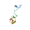
| Title | Structural and functional characterization of an anesthetic binding site in the second cysteine-rich domain of protein kinase C delta | ||||||
 Components Components | Proteine kinase C delta type | ||||||
 Keywords Keywords | METAL BINDING PROTEIN / Proteine kinase C delta / PHOSPHOTRANSFERASE / Anesthetic binding site | ||||||
| Function / homology |  Function and homology information Function and homology informationDAG and IP3 signaling / positive regulation of glucosylceramide catabolic process / positive regulation of sphingomyelin catabolic process / Apoptotic cleavage of cellular proteins / SHC1 events in ERBB2 signaling / VEGFR2 mediated cell proliferation / Effects of PIP2 hydrolysis / regulation of ceramide biosynthetic process / Interferon gamma signaling / HuR (ELAVL1) binds and stabilizes mRNA ...DAG and IP3 signaling / positive regulation of glucosylceramide catabolic process / positive regulation of sphingomyelin catabolic process / Apoptotic cleavage of cellular proteins / SHC1 events in ERBB2 signaling / VEGFR2 mediated cell proliferation / Effects of PIP2 hydrolysis / regulation of ceramide biosynthetic process / Interferon gamma signaling / HuR (ELAVL1) binds and stabilizes mRNA / phospholipase C/protein kinase C signal transduction / TIR domain binding / Calmodulin induced events / protein kinase C signaling / diacylglycerol-dependent, calcium-independent serine/threonine kinase activity / negative regulation of filopodium assembly / endolysosome / positive regulation of ceramide biosynthetic process / Role of phospholipids in phagocytosis / termination of signal transduction / cellular response to hydroperoxide / diacylglycerol-dependent serine/threonine kinase activity / protein kinase C / negative regulation of actin filament polymerization / RHO GTPases Activate NADPH Oxidases / CLEC7A (Dectin-1) signaling / negative regulation of glial cell apoptotic process / collagen metabolic process / neutrophil activation / D-aspartate import across plasma membrane / negative regulation of platelet aggregation / immunoglobulin mediated immune response / B cell proliferation / cellular response to angiotensin / protein tyrosine kinase activator activity / insulin receptor substrate binding / postsynaptic cytosol / positive regulation of superoxide anion generation / negative regulation of MAPK cascade / Neutrophil degranulation / negative regulation of insulin receptor signaling pathway / post-translational protein modification / positive regulation of apoptotic signaling pathway / positive regulation of D-glucose import across plasma membrane / peptidyl-serine phosphorylation / protein serine/threonine kinase binding / regulation of actin cytoskeleton organization / non-specific protein-tyrosine kinase / cell chemotaxis / non-membrane spanning protein tyrosine kinase activity / positive regulation of protein import into nucleus / negative regulation of inflammatory response / cellular senescence / cellular response to hydrogen peroxide / enzyme activator activity / nuclear matrix / cell-cell junction / cellular response to UV / cellular response to oxidative stress / response to oxidative stress / positive regulation of MAPK cascade / protein kinase activity / defense response to bacterium / intracellular signal transduction / positive regulation of apoptotic process / protein serine kinase activity / protein serine/threonine kinase activity / apoptotic process / DNA damage response / protein kinase binding / perinuclear region of cytoplasm / enzyme binding / endoplasmic reticulum / mitochondrion / zinc ion binding / ATP binding / membrane / nucleus / plasma membrane / cytosol / cytoplasm Similarity search - Function | ||||||
| Biological species |  | ||||||
| Method |  X-RAY DIFFRACTION / SYNCHROTRON / MOLECULAR REPLACEMENT / Resolution: 1.357 Å X-RAY DIFFRACTION / SYNCHROTRON / MOLECULAR REPLACEMENT / Resolution: 1.357 Å | ||||||
 Authors Authors | Shanmugasundararaj, S. / Stehle, T. / Miller, K.W. | ||||||
 Citation Citation | Journal: Biophys.J. / Year: 2012 Title: Structural and Functional Characterization of an Anesthetic Binding Site in the Second Cysteine-Rich Domain of Protein Kinase Cdelta Authors: Shanmugasundararaj, S. / Das, J. / Sandberg, W.S. / Zhou, X. / Wang, D. / Messing, R.O. / Bruzik, K.S. / Stehle, T. / Miller, K.W. #1: Journal: Cell(Cambridge,Mass.) / Year: 1995Title: Crystal structure of the cys2 activator binding domain of protein kinase C delta in complex with phorbol ester Authors: Zhang, G. / Kazanietz, M.G. / Blumberg, P.M. / Hurley, J.H. | ||||||
| History |
|
- Structure visualization
| Structure viewer | Molecule: MolmilJmol/JSmol |
|---|
- Downloads & links
-Download
| PDBx/mmCIF format | 3ugl.cif.gz | 77.4 KB | Display | PDBx/mmCIF format |
|---|---|---|---|---|
| PDB format | pdb3ugl.ent.gz | 56.7 KB | Display | PDB format |
| PDBx/mmJSON format | 3ugl.json.gz | Tree view | PDBx/mmJSON format | |
| Others |  Other downloads Other downloads |
-Validation report
| Arichive directory | https://data.pdbj.org/pub/pdb/validation_reports/ug/3uglftp://data.pdbj.org/pub/pdb/validation_reports/ug/3ugl | HTTPS FTP |
|---|
-Related structure data
| Related structure data | 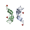 3uejC 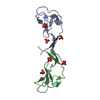 3ueyC  3uffC 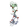 3ugdC  3ugiC 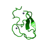 1ptqS S: Starting model for refinement C: citing same article ( |
|---|---|
| Similar structure data |


-Links
 PDBj
PDBj


- Assembly
| Deposited unit | 
| ||||||||
|---|---|---|---|---|---|---|---|---|---|
| 1 |
| ||||||||
| Unit cell |
|
-Components
| #1: Protein | Mass: 7376.554 Da / Num. of mol.: 2 / Fragment: C1B Subdomain of PKC delta, UNP residues 231-280 Source method: isolated from a genetically manipulated source Source: (gene. exp.)  #2: Chemical | ChemComp-ZN /   Mass: 65.409 Da / Num. of mol.: 4 / Source method: obtained synthetically / Formula: Zn Mass: 65.409 Da / Num. of mol.: 4 / Source method: obtained synthetically / Formula: Zn#3: Chemical |   Mass: 72.106 Da / Num. of mol.: 2 / Source method: obtained synthetically / Formula: C4H8O Mass: 72.106 Da / Num. of mol.: 2 / Source method: obtained synthetically / Formula: C4H8O#4: Chemical |   Mass: 94.971 Da / Num. of mol.: 3 / Source method: obtained synthetically / Formula: PO4 Mass: 94.971 Da / Num. of mol.: 3 / Source method: obtained synthetically / Formula: PO4#5: Water | ChemComp-HOH / |  Mass: 18.015 Da / Num. of mol.: 211 / Source method: isolated from a natural source / Formula: H2O Mass: 18.015 Da / Num. of mol.: 211 / Source method: isolated from a natural source / Formula: H2O |
|---|
-Experimental details
-Experiment
| Experiment | Method: X-RAY DIFFRACTION / Number of used crystals: 1 |
|---|
- Sample preparation
| Crystal | Density Matthews: 2.4 Å3/Da / Density % sol: 48.82 % |
|---|---|
| Crystal grow | Temperature: 293 K / Method: vapor diffusion, hanging drop / pH: 7.2 Details: 20% PEG 3350 in 0.2 M ammonium sulfate and 25 mM HEPES pH 7.2. , VAPOR DIFFUSION, HANGING DROP, temperature 293K |
-Data collection
| Diffraction | Mean temperature: 100 K |
|---|---|
| Diffraction source | Source: SYNCHROTRON / Site: NSLS  / Beamline: X6A / Wavelength: 1 Å / Beamline: X6A / Wavelength: 1 Å |
| Detector | Type: ADSC QUANTUM 210 / Detector: CCD / Date: Apr 27, 2004 |
| Radiation | Monochromator: SI(111) CHANNEL CULT MONOCHROMATER / Protocol: SINGLE WAVELENGTH / Monochromatic (M) / Laue (L): M / Scattering type: x-ray |
| Radiation wavelength | Wavelength: 1 Å / Relative weight: 1 |
| Reflection | Resolution: 1.35→10 Å / Num. all: 30362 / Num. obs: 28279 / % possible obs: 92.51 % / Observed criterion σ(F): 4 / Observed criterion σ(I): 2 |
- Processing
| Software |
| |||||||||||||||||||||||||||||||||||||||||||||||||||||||||||||||||||||||||||||||||||||||||||||||||||||||||
|---|---|---|---|---|---|---|---|---|---|---|---|---|---|---|---|---|---|---|---|---|---|---|---|---|---|---|---|---|---|---|---|---|---|---|---|---|---|---|---|---|---|---|---|---|---|---|---|---|---|---|---|---|---|---|---|---|---|---|---|---|---|---|---|---|---|---|---|---|---|---|---|---|---|---|---|---|---|---|---|---|---|---|---|---|---|---|---|---|---|---|---|---|---|---|---|---|---|---|---|---|---|---|---|---|---|---|
| Refinement | Method to determine structure: MOLECULAR REPLACEMENT Starting model: 1PTQ Resolution: 1.357→9.986 Å / SU ML: 0.13 / σ(F): 0 / Phase error: 16.13 / Stereochemistry target values: ML
| |||||||||||||||||||||||||||||||||||||||||||||||||||||||||||||||||||||||||||||||||||||||||||||||||||||||||
| Solvent computation | Shrinkage radii: 0.38 Å / VDW probe radii: 0.7 Å / Solvent model: FLAT BULK SOLVENT MODEL / Bsol: 74.422 Å2 / ksol: 0.546 e/Å3 | |||||||||||||||||||||||||||||||||||||||||||||||||||||||||||||||||||||||||||||||||||||||||||||||||||||||||
| Displacement parameters |
| |||||||||||||||||||||||||||||||||||||||||||||||||||||||||||||||||||||||||||||||||||||||||||||||||||||||||
| Refinement step | Cycle: LAST / Resolution: 1.357→9.986 Å
| |||||||||||||||||||||||||||||||||||||||||||||||||||||||||||||||||||||||||||||||||||||||||||||||||||||||||
| Refine LS restraints |
| |||||||||||||||||||||||||||||||||||||||||||||||||||||||||||||||||||||||||||||||||||||||||||||||||||||||||
| LS refinement shell |
|
