 Movie
Movie Controller


+ Open data

- Basic information
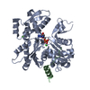
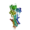
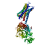
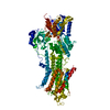
| Entry | Database: PDB / ID: 3ue5 | ||||||
|---|---|---|---|---|---|---|---|
| Title | ECP-cleaved Actin in complex with Spir domain D | ||||||
 Components Components |
| ||||||
 Keywords Keywords | CONTRACTILE PROTEIN/TRANSPORT PROTEIN / CONTRACTILE PROTEIN / CONTRACTILE PROTEIN-TRANSPORT PROTEIN complex | ||||||
| Function / homology |  Function and homology information Function and homology informationchorion-containing eggshell formation / pole plasm RNA localization / oocyte karyosome formation / establishment of meiotic spindle localization / actin filament-based process / pole plasm assembly / actin filament network formation / polar body extrusion after meiotic divisions / pole plasm oskar mRNA localization / actin nucleation ...chorion-containing eggshell formation / pole plasm RNA localization / oocyte karyosome formation / establishment of meiotic spindle localization / actin filament-based process / pole plasm assembly / actin filament network formation / polar body extrusion after meiotic divisions / pole plasm oskar mRNA localization / actin nucleation / Golgi vesicle transport / cleavage furrow formation / positive regulation of mitochondrial fission / cytoskeletal motor activator activity / oogenesis / myosin heavy chain binding / regulation of cytoskeleton organization / tropomyosin binding / actin filament bundle / troponin I binding / filamentous actin / mesenchyme migration / skeletal muscle myofibril / actin filament bundle assembly / striated muscle thin filament / skeletal muscle thin filament assembly / actin monomer binding / skeletal muscle fiber development / vesicle-mediated transport / stress fiber / titin binding / actin filament polymerization / cytoplasmic vesicle membrane / actin filament organization / filopodium / actin filament / Hydrolases; Acting on acid anhydrides; Acting on acid anhydrides to facilitate cellular and subcellular movement / calcium-dependent protein binding / protein transport / lamellipodium / actin binding / actin cytoskeleton organization / cell body / cell cortex / microtubule binding / cytoskeleton / mitochondrial outer membrane / protein domain specific binding / hydrolase activity / calcium ion binding / positive regulation of gene expression / perinuclear region of cytoplasm / magnesium ion binding / ATP binding / identical protein binding / plasma membrane / cytoplasm Similarity search - Function | ||||||
| Biological species |   | ||||||
| Method |  X-RAY DIFFRACTION / SYNCHROTRON / MOLECULAR REPLACEMENT / molecular replacement / Resolution: 2.76 Å X-RAY DIFFRACTION / SYNCHROTRON / MOLECULAR REPLACEMENT / molecular replacement / Resolution: 2.76 Å | ||||||
 Authors Authors | Chen, C. / Phillips, M. / Sawaya, M.R. / Ralston, C.Y. / Quinlan, M.E. | ||||||
 Citation Citation | Journal: J.Biol.Chem. / Year: 2012 Title: Multiple Forms of Spire-Actin Complexes and their Functional Consequences. Authors: Chen, C.K. / Sawaya, M.R. / Phillips, M.L. / Reisler, E. / Quinlan, M.E. | ||||||
| History |
|
- Structure visualization
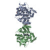
| Structure viewer | Molecule: MolmilJmol/JSmol |
|---|
- Downloads & links
-Download
| PDBx/mmCIF format | 3ue5.cif.gz | 170.7 KB | Display | PDBx/mmCIF format |
|---|---|---|---|---|
| PDB format | pdb3ue5.ent.gz | 132.8 KB | Display | PDB format |
| PDBx/mmJSON format | 3ue5.json.gz | Tree view | PDBx/mmJSON format | |
| Others |  Other downloads Other downloads |
-Validation report
| Arichive directory | https://data.pdbj.org/pub/pdb/validation_reports/ue/3ue5ftp://data.pdbj.org/pub/pdb/validation_reports/ue/3ue5 | HTTPS FTP |
|---|
-Related structure data

-Links
 PDBj
PDBj


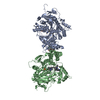
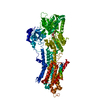
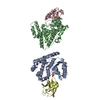
- Assembly
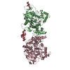
| Deposited unit | 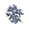
| ||||||||
|---|---|---|---|---|---|---|---|---|---|
| 1 | 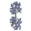
| ||||||||
| 2 | 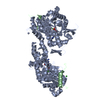
| ||||||||
| 3 | 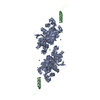
| ||||||||
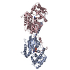
| Unit cell |
| ||||||||
| Components on special symmetry positions |
|
-Components
-Protein , 2 types, 2 molecules AB
| #1: Protein | Mass: 41862.613 Da / Num. of mol.: 1 / Source method: isolated from a natural source / Details: skeletal / Source: (natural) |
|---|---|
| #2: Protein | Mass: 7320.585 Da / Num. of mol.: 1 / Fragment: UNP residues 428-485 Source method: isolated from a genetically manipulated source Source: (gene. exp.)  |
-Non-polymers , 4 types, 44 molecules 


| #3: Chemical | ChemComp-CA /  Mass: 40.078 Da / Num. of mol.: 1 / Source method: obtained synthetically / Formula: Ca Mass: 40.078 Da / Num. of mol.: 1 / Source method: obtained synthetically / Formula: Ca | ||
|---|---|---|---|
| #4: Chemical | ChemComp-ATP /  Mass: 507.181 Da / Num. of mol.: 1 / Source method: obtained synthetically / Formula: C10H16N5O13P3 / Comment: ATP, energy-carrying molecule*YM Mass: 507.181 Da / Num. of mol.: 1 / Source method: obtained synthetically / Formula: C10H16N5O13P3 / Comment: ATP, energy-carrying molecule*YM | ||
| #5: Chemical |  Mass: 35.453 Da / Num. of mol.: 2 / Source method: obtained synthetically / Formula: Cl Mass: 35.453 Da / Num. of mol.: 2 / Source method: obtained synthetically / Formula: Cl#6: Water | ChemComp-HOH / | Mass: 18.015 Da / Num. of mol.: 40 / Source method: isolated from a natural source / Formula: H2O |
-Details
| Has protein modification | Y |
|---|
-Experimental details
-Experiment
| Experiment | Method: X-RAY DIFFRACTION / Number of used crystals: 1 |
|---|
- Sample preparation
| Crystal | Density Matthews: 2.32 Å3/Da / Density % sol: 46.99 % |
|---|---|
| Crystal grow | Temperature: 298 K / Method: vapor diffusion, hanging drop / pH: 8.5 Details: Tris pH 8.5, magnesium chloride, PEG-8000, vapor diffusion, hanging drop, temperature 298K |
-Data collection
| Diffraction | Mean temperature: 100 K | |||||||||||||||||||||||||||||||||||||||||||||||||||||||||||||||||||||||||||||
|---|---|---|---|---|---|---|---|---|---|---|---|---|---|---|---|---|---|---|---|---|---|---|---|---|---|---|---|---|---|---|---|---|---|---|---|---|---|---|---|---|---|---|---|---|---|---|---|---|---|---|---|---|---|---|---|---|---|---|---|---|---|---|---|---|---|---|---|---|---|---|---|---|---|---|---|---|---|---|
| Diffraction source | Source: SYNCHROTRON / Site: ALS  / Beamline: 8.2.1 / Wavelength: 1 Å / Beamline: 8.2.1 / Wavelength: 1 Å | |||||||||||||||||||||||||||||||||||||||||||||||||||||||||||||||||||||||||||||
| Detector | Type: ADSC QUANTUM 315 / Detector: CCD / Date: Feb 5, 2008 | |||||||||||||||||||||||||||||||||||||||||||||||||||||||||||||||||||||||||||||
| Radiation | Protocol: SINGLE WAVELENGTH / Monochromatic (M) / Laue (L): M / Scattering type: x-ray | |||||||||||||||||||||||||||||||||||||||||||||||||||||||||||||||||||||||||||||
| Radiation wavelength | Wavelength: 1 Å / Relative weight: 1 | |||||||||||||||||||||||||||||||||||||||||||||||||||||||||||||||||||||||||||||
| Reflection | Resolution: 2.76→90 Å / Num. all: 10542 / Num. obs: 10542 / % possible obs: 89.6 % / Observed criterion σ(I): -3 / Redundancy: 2.7 % / Biso Wilson estimate: 46 Å2 / Rmerge(I) obs: 0.105 / Χ2: 0.963 / Net I/σ(I): 8.8 | |||||||||||||||||||||||||||||||||||||||||||||||||||||||||||||||||||||||||||||
| Reflection shell |
|
-Phasing
| Phasing | Method: molecular replacement | |||||||||
|---|---|---|---|---|---|---|---|---|---|---|
| Phasing MR | Model details: Phaser MODE: MR_AUTO
|
- Processing
| Software |
| ||||||||||||||||||||||||||||||||||||||||||||||||||||||||||||||||||||||||||||||||||||||||||||||||||||||||||||
|---|---|---|---|---|---|---|---|---|---|---|---|---|---|---|---|---|---|---|---|---|---|---|---|---|---|---|---|---|---|---|---|---|---|---|---|---|---|---|---|---|---|---|---|---|---|---|---|---|---|---|---|---|---|---|---|---|---|---|---|---|---|---|---|---|---|---|---|---|---|---|---|---|---|---|---|---|---|---|---|---|---|---|---|---|---|---|---|---|---|---|---|---|---|---|---|---|---|---|---|---|---|---|---|---|---|---|---|---|---|
| Refinement | Method to determine structure: MOLECULAR REPLACEMENT / Resolution: 2.76→50.24 Å / Cor.coef. Fo:Fc: 0.8964 / Cor.coef. Fo:Fc free: 0.8718 / Occupancy max: 1 / Occupancy min: 1 / Cross valid method: THROUGHOUT / σ(F): 0 / Stereochemistry target values: Engh & Huber
| ||||||||||||||||||||||||||||||||||||||||||||||||||||||||||||||||||||||||||||||||||||||||||||||||||||||||||||
| Displacement parameters | Biso max: 149.31 Å2 / Biso mean: 48.4404 Å2 / Biso min: 15.64 Å2
| ||||||||||||||||||||||||||||||||||||||||||||||||||||||||||||||||||||||||||||||||||||||||||||||||||||||||||||
| Refine analyze | Luzzati coordinate error obs: 0.364 Å | ||||||||||||||||||||||||||||||||||||||||||||||||||||||||||||||||||||||||||||||||||||||||||||||||||||||||||||
| Refinement step | Cycle: LAST / Resolution: 2.76→50.24 Å
| ||||||||||||||||||||||||||||||||||||||||||||||||||||||||||||||||||||||||||||||||||||||||||||||||||||||||||||
| Refine LS restraints |
| ||||||||||||||||||||||||||||||||||||||||||||||||||||||||||||||||||||||||||||||||||||||||||||||||||||||||||||
| LS refinement shell | Resolution: 2.76→3.08 Å / Total num. of bins used: 5
| ||||||||||||||||||||||||||||||||||||||||||||||||||||||||||||||||||||||||||||||||||||||||||||||||||||||||||||
| Refinement TLS params. | Method: refined / Refine-ID: X-RAY DIFFRACTION
| ||||||||||||||||||||||||||||||||||||||||||||||||||||||||||||||||||||||||||||||||||||||||||||||||||||||||||||
| Refinement TLS group |
|
