 Movie
Movie Controller

[English] 日本語
 Yorodumi
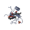
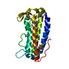
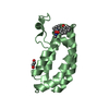
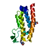
- PDB-3u99: The experimental X-ray structure of the new diheme cytochrome typ... -
+ Open data

- Basic information
| Entry | Database: PDB / ID: 3u99 | ||||||
|---|---|---|---|---|---|---|---|
| Title | The experimental X-ray structure of the new diheme cytochrome type c from Shewanella baltica OS155 sb-DHC | ||||||
 Components Components | Diheme cytochrome c | ||||||
 Keywords Keywords | ELECTRON TRANSPORT / Cytochrome c fold / Electron transfer protein / diheme protein / bacterium Shewanella Baltica OS155 | ||||||
| Function / homology | Dihaem cytochrome c / Dihaem cytochrome c / metal ion binding / HEME C / Diheme cytochrome c Function and homology information Function and homology information | ||||||
| Biological species |  Shewanella baltica (bacteria) Shewanella baltica (bacteria) | ||||||
| Method |  X-RAY DIFFRACTION / SYNCHROTRON / MOLECULAR REPLACEMENT / Resolution: 1.146 Å X-RAY DIFFRACTION / SYNCHROTRON / MOLECULAR REPLACEMENT / Resolution: 1.146 Å | ||||||
 Authors Authors | De March, M. / Di Rocco, G. / Geremia, S. | ||||||
 Citation Citation | Journal: J.Biomol.Struct.Dyn. / Year: 2015 Title: High-resolution crystal structure of the recombinant diheme cytochrome c from Shewanella baltica (OS155). Authors: De March, M. / Di Rocco, G. / Hickey, N. / Geremia, S. #1: Journal: J.Biol.Inorg.Chem. / Year: 2011 Title: Cloning, expression, and physicochemical characterization of a new diheme cytochrome c from Shewanella baltica OS155. Authors: Di Rocco, G. / Battistuzzi, G. / Bortolotti, C.A. / Borsari, M. / Ferrari, E. / Monari, S. / Sola, M. #2: Journal: Biochemistry / Year: 2006Title: Structural and functional studies on DHC, the diheme cytochrome c from Rhodobacter sphaeroides, and its interaction with SHP, the sphaeroides heme protein. Authors: Gibson, H.R. / Mowat, C.G. / Miles, C.S. / Li, B.R. / Leys, D. / Reid, G.A. / Chapman, S.K. #3: Journal: Biochemistry / Year: 2005Title: Structural and biochemical characterization of DHC2, a novel diheme cytochrome c from Geobacter sulfurreducens. Authors: Heitmann, D. / Einsle, O. #4: Journal: Biochemistry / Year: 2002Title: The 1.25 A resolution structure of the diheme NapB subunit of soluble nitrate reductase reveals a novel cytochrome c fold with a stacked heme arrangement. Authors: Brige, A. / Leys, D. / Meyer, T.E. / Cusanovich, M.A. / Van Beeumen, J.J. | ||||||
| History |
|
- Structure visualization
| Structure viewer | Molecule: MolmilJmol/JSmol |
|---|
- Downloads & links
-Download
| PDBx/mmCIF format | 3u99.cif.gz | 102.7 KB | Display | PDBx/mmCIF format |
|---|---|---|---|---|
| PDB format | pdb3u99.ent.gz | 80.4 KB | Display | PDB format |
| PDBx/mmJSON format | 3u99.json.gz | Tree view | PDBx/mmJSON format | |
| Others |  Other downloads Other downloads |
-Validation report
| Arichive directory | https://data.pdbj.org/pub/pdb/validation_reports/u9/3u99ftp://data.pdbj.org/pub/pdb/validation_reports/u9/3u99 | HTTPS FTP |
|---|
-Related structure data
| Related structure data |  1fw5S S: Starting model for refinement |
|---|---|
| Similar structure data |


-Links
 PDBj
PDBj


- Assembly
| Deposited unit | 
| ||||||||
|---|---|---|---|---|---|---|---|---|---|
| 1 |
| ||||||||
| Unit cell |
|
-Components
| #1: Protein | Mass: 16388.250 Da / Num. of mol.: 1 / Fragment: UNP Residues 40-187 Source method: isolated from a genetically manipulated source Source: (gene. exp.) Shewanella baltica (bacteria) / Strain: OS155 / ATCC BAA-1091 / Gene: Sbal_0241 / Production host: | ||||
|---|---|---|---|---|---|
| #2: Chemical |   Mass: 618.503 Da / Num. of mol.: 2 / Source method: obtained synthetically / Formula: C34H34FeN4O4 Mass: 618.503 Da / Num. of mol.: 2 / Source method: obtained synthetically / Formula: C34H34FeN4O4#3: Water | ChemComp-HOH / |  Mass: 18.015 Da / Num. of mol.: 250 / Source method: isolated from a natural source / Formula: H2O Mass: 18.015 Da / Num. of mol.: 250 / Source method: isolated from a natural source / Formula: H2OHas protein modification | Y | |
-Experimental details
-Experiment
| Experiment | Method: X-RAY DIFFRACTION / Number of used crystals: 1 |
|---|
- Sample preparation
| Crystal | Density Matthews: 2.17 Å3/Da / Density % sol: 43.28 % |
|---|---|
| Crystal grow | Temperature: 298 K / Method: vapor diffusion, hanging drop / pH: 6.5 Details: Tris pH 6.5, PEG3350, Ammonium Sulphate, Dithionite, VAPOR DIFFUSION, HANGING DROP, temperature 298K |
-Data collection
| Diffraction | Mean temperature: 100 K |
|---|---|
| Diffraction source | Source: SYNCHROTRON / Site: ELETTRA  / Beamline: 5.2R / Wavelength: 1 Å / Beamline: 5.2R / Wavelength: 1 Å |
| Detector | Type: PSI PILATUS 6M / Detector: PIXEL / Date: Dec 19, 2010 |
| Radiation | Monochromator: GRAPHITE / Protocol: SINGLE WAVELENGTH / Monochromatic (M) / Laue (L): M / Scattering type: x-ray |
| Radiation wavelength | Wavelength: 1 Å / Relative weight: 1 |
| Reflection | Resolution: 1.08→17.97 Å / Num. all: 58315 / Num. obs: 58150 / % possible obs: 96.9 % / Observed criterion σ(F): 2 / Observed criterion σ(I): 2 / Rmerge(I) obs: 0.107 |
| Reflection shell | Resolution: 1.08→2.45 Å / % possible all: 96.9 |
- Processing
| Software |
| ||||||||||||||||||||
|---|---|---|---|---|---|---|---|---|---|---|---|---|---|---|---|---|---|---|---|---|---|
| Refinement | Method to determine structure: MOLECULAR REPLACEMENT Starting model: PDB ENTRY 1FW5 Resolution: 1.146→10.8 Å / σ(F): 2 / Stereochemistry target values: Engh & Huber
| ||||||||||||||||||||
| Refinement step | Cycle: LAST / Resolution: 1.146→10.8 Å
| ||||||||||||||||||||
| Refine LS restraints |
|
