 Movie
Movie Controller

[English] 日本語
 Yorodumi
- PDB-1jni: Structure of the NapB subunit of the periplasmic nitrate reductas... -
+ Open data

- Basic information
| Entry | Database: PDB / ID: 1jni | |||||||||
|---|---|---|---|---|---|---|---|---|---|---|
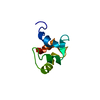
| Title | Structure of the NapB subunit of the periplasmic nitrate reductase from Haemophilus influenzae. | |||||||||
 Components Components | DIHEME CYTOCHROME C NAPB | |||||||||
 Keywords Keywords | OXIDOREDUCTASE / dihaem cytochrome c / proteolytic fragment / nitrate reductase subunit | |||||||||
| Function / homology |  Function and homology information Function and homology informationanaerobic respiration / molecular adaptor activity / periplasmic space / metal ion binding Similarity search - Function | |||||||||
| Biological species |  Haemophilus influenzae (bacteria) Haemophilus influenzae (bacteria) | |||||||||
| Method |  X-RAY DIFFRACTION / SYNCHROTRON / MAD / Resolution: 1.25 Å X-RAY DIFFRACTION / SYNCHROTRON / MAD / Resolution: 1.25 Å | |||||||||
 Authors Authors | Brige, A. / Leys, D. / Meyer, T.E. / Cusanovich, M.A. / Van Beeumen, J.J. | |||||||||
 Citation Citation | Journal: Biochemistry / Year: 2002 Title: The 1.25 A resolution structure of the diheme NapB subunit of soluble nitrate reductase reveals a novel cytochrome c fold with a stacked heme arrangement. Authors: Brige, A. / Leys, D. / Meyer, T.E. / Cusanovich, M.A. / Van Beeumen, J.J. | |||||||||
| History |
|
- Structure visualization
| Structure viewer | Molecule: MolmilJmol/JSmol |
|---|
- Downloads & links
-Download
| PDBx/mmCIF format | 1jni.cif.gz | 50.5 KB | Display | PDBx/mmCIF format |
|---|---|---|---|---|
| PDB format | pdb1jni.ent.gz | 34.2 KB | Display | PDB format |
| PDBx/mmJSON format | 1jni.json.gz | Tree view | PDBx/mmJSON format | |
| Others |  Other downloads Other downloads |
-Validation report
| Arichive directory | https://data.pdbj.org/pub/pdb/validation_reports/jn/1jniftp://data.pdbj.org/pub/pdb/validation_reports/jn/1jni | HTTPS FTP |
|---|
-Related structure data
| Similar structure data |
|---|


-Links
 PDBj
PDBj


- Assembly
| Deposited unit | 
| ||||||||||||
|---|---|---|---|---|---|---|---|---|---|---|---|---|---|
| 1 |
| ||||||||||||
| Unit cell |
| ||||||||||||
| Components on special symmetry positions |
|
-Components
| #1: Protein | Mass: 13444.035 Da / Num. of mol.: 1 Fragment: SMALL SUBUNIT OF THE PERIPLASMIC NITRATE REDUCTASE Source method: isolated from a genetically manipulated source Source: (gene. exp.) Haemophilus influenzae (bacteria) / Production host: | ||||
|---|---|---|---|---|---|
| #2: Chemical |   Mass: 618.503 Da / Num. of mol.: 2 / Source method: obtained synthetically / Formula: C34H34FeN4O4 Mass: 618.503 Da / Num. of mol.: 2 / Source method: obtained synthetically / Formula: C34H34FeN4O4#3: Water | ChemComp-HOH / |  Mass: 18.015 Da / Num. of mol.: 128 / Source method: isolated from a natural source / Formula: H2O Mass: 18.015 Da / Num. of mol.: 128 / Source method: isolated from a natural source / Formula: H2OHas protein modification | Y | |
-Experimental details
-Experiment
| Experiment | Method: X-RAY DIFFRACTION / Number of used crystals: 1 |
|---|
- Sample preparation
| Crystal | Density Matthews: 1.67 Å3/Da / Density % sol: 26.57 % | ||||||||||||||||||||||||||||||||||||||||||
|---|---|---|---|---|---|---|---|---|---|---|---|---|---|---|---|---|---|---|---|---|---|---|---|---|---|---|---|---|---|---|---|---|---|---|---|---|---|---|---|---|---|---|---|
| Crystal grow | Temperature: 296 K / Method: vapor diffusion, hanging drop / pH: 5.5 Details: ammonium sulphate, pH 5.5, VAPOR DIFFUSION, HANGING DROP at 296K | ||||||||||||||||||||||||||||||||||||||||||
| Crystal grow | *PLUS Temperature: 277 K / pH: 6.5 / Details: Brige, A., (2001) Acta Crystallogr, D57, 418. | ||||||||||||||||||||||||||||||||||||||||||
| Components of the solutions | *PLUS
|
-Data collection
| Diffraction | Mean temperature: 173 K |
|---|---|
| Diffraction source | Source: SYNCHROTRON / Site: ESRF  / Beamline: ID13 / Wavelength: 0.964 Å / Beamline: ID13 / Wavelength: 0.964 Å |
| Detector | Type: MARRESEARCH / Detector: CCD / Date: Aug 14, 2000 |
| Radiation | Monochromator: SAGITALLY FOCUSED Si(111) / Protocol: MAD / Monochromatic (M) / Laue (L): M / Scattering type: x-ray |
| Radiation wavelength | Wavelength: 0.964 Å / Relative weight: 1 |
| Reflection | Resolution: 1.24→10 Å / Num. obs: 22337 / % possible obs: 84.4 % / Observed criterion σ(F): 0 / Observed criterion σ(I): 0 / Biso Wilson estimate: 28 Å2 / Rmerge(I) obs: 0.089 / Net I/σ(I): 17 |
| Reflection shell | Resolution: 1.24→1.3 Å / Rmerge(I) obs: 0.316 / Mean I/σ(I) obs: 1.37 / % possible all: 65.5 |
| Reflection | *PLUS Highest resolution: 1.25 Å / Lowest resolution: 10 Å / Num. measured all: 214619 / Rmerge(I) obs: 0.089 |
| Reflection shell | *PLUS Highest resolution: 1.25 Å / Lowest resolution: 1.26 Å / % possible obs: 65.47 % / Rmerge(I) obs: 0.316 |
- Processing
| Software |
| |||||||||||||||||||||||||
|---|---|---|---|---|---|---|---|---|---|---|---|---|---|---|---|---|---|---|---|---|---|---|---|---|---|---|
| Refinement | Method to determine structure: MAD / Resolution: 1.25→10 Å / Isotropic thermal model: anisotropic model / σ(F): 0 / σ(I): 0 / Stereochemistry target values: Engh & Huber
| |||||||||||||||||||||||||
| Displacement parameters | Biso mean: 26.855 Å2 | |||||||||||||||||||||||||
| Refinement step | Cycle: LAST / Resolution: 1.25→10 Å
| |||||||||||||||||||||||||
| Refine LS restraints |
| |||||||||||||||||||||||||
| Software | *PLUS Name: SHELXL / Version: 97 / Classification: refinement | |||||||||||||||||||||||||
| Refinement | *PLUS Lowest resolution: 10 Å / Rfactor all: 0.16 / Rfactor obs: 0.1596 / Rfactor Rfree: 0.2165 / Rfactor Rwork: 0.1596 | |||||||||||||||||||||||||
| Solvent computation | *PLUS | |||||||||||||||||||||||||
| Displacement parameters | *PLUS | |||||||||||||||||||||||||
| Refine LS restraints | *PLUS
|
