 Movie
Movie Controller

[English] 日本語
 Yorodumi
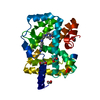
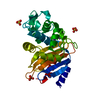
- PDB-3u6g: Crystal structure of a domain of unknown function, DUF4425 (BVU_3... -
+ Open data

- Basic information
| Entry | Database: PDB / ID: 3u6g | ||||||
|---|---|---|---|---|---|---|---|
| Title | Crystal structure of a domain of unknown function, DUF4425 (BVU_3708) from Bacteroides vulgatus ATCC 8482 at 1.35 A resolution | ||||||
 Components Components | uncharacterized protein DUF4425 | ||||||
 Keywords Keywords | Structural Genomics / Unknown Function / DUF4425 / immunoglobulin-like fold / Joint Center for Structural Genomics / JCSG / Protein Structure Initiative / PSI-BIOLOGY | ||||||
| Function / homology | Lipoxygenase-1 - #60 / PLAT/LH2 and C2-like Ca2+-binding lipoprotein / PLAT/LH2 and C2-like Ca2+-binding lipoprotein / Lipoxygenase-1 / Prokaryotic membrane lipoprotein lipid attachment site profile. / Sandwich / Mainly Beta / Lipoprotein Function and homology information Function and homology information | ||||||
| Biological species |  Bacteroides vulgatus (bacteria) Bacteroides vulgatus (bacteria) | ||||||
| Method |  X-RAY DIFFRACTION / SYNCHROTRON / MAD / Resolution: 1.35 Å X-RAY DIFFRACTION / SYNCHROTRON / MAD / Resolution: 1.35 Å | ||||||
 Authors Authors | Joint Center for Structural Genomics (JCSG) | ||||||
 Citation Citation | Journal: To be published Title: Crystal structure of a domain of unknown function, DUF4425 (BVU_3708) from Bacteroides vulgatus ATCC 8482 at 1.35 A resolution Authors: Joint Center for Structural Genomics (JCSG) | ||||||
| History |
|
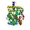
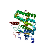
- Structure visualization
| Structure viewer | Molecule: MolmilJmol/JSmol |
|---|
- Downloads & links
-Download
| PDBx/mmCIF format | 3u6g.cif.gz | 118.6 KB | Display | PDBx/mmCIF format |
|---|---|---|---|---|
| PDB format | pdb3u6g.ent.gz | 95.9 KB | Display | PDB format |
| PDBx/mmJSON format | 3u6g.json.gz | Tree view | PDBx/mmJSON format | |
| Others |  Other downloads Other downloads |
-Validation report
| Summary document | 3u6g_validation.pdf.gz | 431.1 KB | Display | wwPDB validaton report |
|---|---|---|---|---|
| Full document | 3u6g_full_validation.pdf.gz | 434.4 KB | Display | |
| Data in XML | 3u6g_validation.xml.gz | 14.1 KB | Display | |
| Data in CIF | 3u6g_validation.cif.gz | 20.4 KB | Display | |
| Arichive directory | https://data.pdbj.org/pub/pdb/validation_reports/u6/3u6gftp://data.pdbj.org/pub/pdb/validation_reports/u6/3u6g | HTTPS FTP |
-Related structure data
| Similar structure data | |
|---|---|
| Other databases |


-Links
 PDBj
PDBj


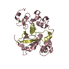
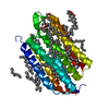
- Assembly
| Deposited unit | 
| ||||||||
|---|---|---|---|---|---|---|---|---|---|
| 1 |
| ||||||||
| Unit cell |
| ||||||||
| Components on special symmetry positions |
| ||||||||
| Details | ANALYTICAL SIZE EXCLUSION CHROMATOGRAPHY SUPPORTS THE ASSIGNMENT OF A DIMER AS A SIGNIFICANT OLIGOMERIZATION STATE IN SOLUTION. |
-Components
| #1: Protein | Mass: 13581.699 Da / Num. of mol.: 2 Source method: isolated from a genetically manipulated source Source: (gene. exp.) Bacteroides vulgatus (bacteria) / Strain: ATCC 8482 / Gene: BVU_3708 / Plasmid: SpeedET / Production host: #2: Water | ChemComp-HOH / |  Mass: 18.015 Da / Num. of mol.: 244 / Source method: isolated from a natural source / Formula: H2O Mass: 18.015 Da / Num. of mol.: 244 / Source method: isolated from a natural source / Formula: H2OSequence details | THE CONSTRUCT WAS EXPRESSED WITH A PURIFICATION TAG MGSDKIHHHHHHENLYFQG. THE TAG WAS REMOVED WITH ...THE CONSTRUCT WAS EXPRESSED WITH A PURIFICATI | |
|---|
-Experimental details
-Experiment
| Experiment | Method: X-RAY DIFFRACTION / Number of used crystals: 1 |
|---|
- Sample preparation
| Crystal | Density Matthews: 1.96 Å3/Da / Density % sol: 37.29 % |
|---|---|
| Crystal grow | Temperature: 293 K / Method: vapor diffusion, sitting drop / pH: 7.5 Details: 0.1M HEPES pH 7.5, 1.4M tri-sodium citrate, NANODROP, VAPOR DIFFUSION, SITTING DROP, temperature 293K |
-Data collection
| Diffraction | Mean temperature: 100 K | |||||||||||||||||||||||||||||||||||||||||||||||||||||||||||||||||||||||||||||
|---|---|---|---|---|---|---|---|---|---|---|---|---|---|---|---|---|---|---|---|---|---|---|---|---|---|---|---|---|---|---|---|---|---|---|---|---|---|---|---|---|---|---|---|---|---|---|---|---|---|---|---|---|---|---|---|---|---|---|---|---|---|---|---|---|---|---|---|---|---|---|---|---|---|---|---|---|---|---|
| Diffraction source | Source: SYNCHROTRON / Site: SSRL  / Beamline: BL14-1 / Wavelength: 0.96109,0.97961 / Beamline: BL14-1 / Wavelength: 0.96109,0.97961 | |||||||||||||||||||||||||||||||||||||||||||||||||||||||||||||||||||||||||||||
| Detector | Type: MARMOSAIC 325 mm CCD / Detector: CCD / Date: Jul 18, 2011 Details: Vertical focusing mirror; double crystal Si(111) monochromator | |||||||||||||||||||||||||||||||||||||||||||||||||||||||||||||||||||||||||||||
| Radiation | Monochromator: double crystal Si(111) / Protocol: MAD / Monochromatic (M) / Laue (L): M / Scattering type: x-ray | |||||||||||||||||||||||||||||||||||||||||||||||||||||||||||||||||||||||||||||
| Radiation wavelength |
| |||||||||||||||||||||||||||||||||||||||||||||||||||||||||||||||||||||||||||||
| Reflection | Resolution: 1.35→27.96 Å / Num. obs: 45712 / % possible obs: 93.6 % / Observed criterion σ(I): -3 / Biso Wilson estimate: 19.297 Å2 / Rmerge(I) obs: 0.027 / Net I/σ(I): 14.43 | |||||||||||||||||||||||||||||||||||||||||||||||||||||||||||||||||||||||||||||
| Reflection shell | Diffraction-ID: 1
|
-Phasing
| Phasing | Method: MAD |
|---|
- Processing
| Software |
| |||||||||||||||||||||||||||||||||||||||||||||||||||||||||||||||||||||||||||
|---|---|---|---|---|---|---|---|---|---|---|---|---|---|---|---|---|---|---|---|---|---|---|---|---|---|---|---|---|---|---|---|---|---|---|---|---|---|---|---|---|---|---|---|---|---|---|---|---|---|---|---|---|---|---|---|---|---|---|---|---|---|---|---|---|---|---|---|---|---|---|---|---|---|---|---|---|
| Refinement | Method to determine structure: MAD / Resolution: 1.35→27.96 Å / Cor.coef. Fo:Fc: 0.972 / Cor.coef. Fo:Fc free: 0.956 / Occupancy max: 1 / Occupancy min: 0.37 / SU B: 2.236 / SU ML: 0.041 / Cross valid method: THROUGHOUT / σ(F): 0 / ESU R Free: 0.066 Stereochemistry target values: MAXIMUM LIKELIHOOD WITH PHASES Details: 1. A MET-INHIBITION PROTOCOL WAS USED FOR SELENOMETHIONINE INCORPORATION DURING PROTEIN EXPRESSION. THE OCCUPANCY OF THE SE ATOMS IN THE MSE RESIDUES WAS REDUCED TO 0.75 FOR THE REDUCED ...Details: 1. A MET-INHIBITION PROTOCOL WAS USED FOR SELENOMETHIONINE INCORPORATION DURING PROTEIN EXPRESSION. THE OCCUPANCY OF THE SE ATOMS IN THE MSE RESIDUES WAS REDUCED TO 0.75 FOR THE REDUCED SCATTERING POWER DUE TO PARTIAL S-MET INCORPORATION. 2. HYDROGENS HAVE BEEN ADDED IN THE RIDING POSITIONS.
| |||||||||||||||||||||||||||||||||||||||||||||||||||||||||||||||||||||||||||
| Solvent computation | Ion probe radii: 0.8 Å / Shrinkage radii: 0.8 Å / VDW probe radii: 1.2 Å / Solvent model: MASK | |||||||||||||||||||||||||||||||||||||||||||||||||||||||||||||||||||||||||||
| Displacement parameters | Biso max: 60.6 Å2 / Biso mean: 23.0605 Å2 / Biso min: 8.96 Å2
| |||||||||||||||||||||||||||||||||||||||||||||||||||||||||||||||||||||||||||
| Refinement step | Cycle: LAST / Resolution: 1.35→27.96 Å
| |||||||||||||||||||||||||||||||||||||||||||||||||||||||||||||||||||||||||||
| Refine LS restraints |
| |||||||||||||||||||||||||||||||||||||||||||||||||||||||||||||||||||||||||||
| LS refinement shell | Resolution: 1.351→1.386 Å / Total num. of bins used: 20
|
