 Movie
Movie Controller

[English] 日本語
 Yorodumi
- PDB-3u3f: Structural basis for the interaction of Pyk2 PAT domain with paxi... -
+ Open data

- Basic information
| Entry | Database: PDB / ID: 3u3f | ||||||
|---|---|---|---|---|---|---|---|
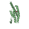
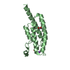
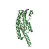
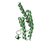
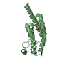
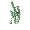
| Title | Structural basis for the interaction of Pyk2 PAT domain with paxillin LD motifs | ||||||
 Components Components |
| ||||||
 Keywords Keywords | TRANSFERASE/signaling protein / 4-helix bundle / focal adhesion / tyrosine kinase / paxillin / TRANSFERASE-signaling protein complex | ||||||
| Function / homology |  Function and homology information Function and homology informationregulation of macrophage chemotaxis / neurotransmitter receptor regulator activity / positive regulation of B cell chemotaxis / marginal zone B cell differentiation / Regulation of cytoskeletal remodeling and cell spreading by IPP complex components / Localization of the PINCH-ILK-PARVIN complex to focal adhesions / regulation of ubiquitin-dependent protein catabolic process / endothelin receptor signaling pathway / negative regulation of myeloid cell differentiation / Regulation of MITF-M-dependent genes involved in extracellular matrix, focal adhesion and epithelial-to-mesenchymal transition ...regulation of macrophage chemotaxis / neurotransmitter receptor regulator activity / positive regulation of B cell chemotaxis / marginal zone B cell differentiation / Regulation of cytoskeletal remodeling and cell spreading by IPP complex components / Localization of the PINCH-ILK-PARVIN complex to focal adhesions / regulation of ubiquitin-dependent protein catabolic process / endothelin receptor signaling pathway / negative regulation of myeloid cell differentiation / Regulation of MITF-M-dependent genes involved in extracellular matrix, focal adhesion and epithelial-to-mesenchymal transition / neuropilin binding / regulation of postsynaptic density assembly / vinculin binding / activation of Janus kinase activity / negative regulation of bone mineralization / cortical cytoskeleton organization / cellular response to fluid shear stress / peptidyl-tyrosine autophosphorylation / signal complex assembly / calcium/calmodulin-dependent protein kinase activity / apical dendrite / Interleukin-2 signaling / sprouting angiogenesis / regulation of release of sequestered calcium ion into cytosol / Signal regulatory protein family interactions / positive regulation of protein kinase activity / microtubule associated complex / growth hormone receptor signaling pathway / positive regulation of ubiquitin-dependent protein catabolic process / NMDA selective glutamate receptor complex / positive regulation of cell-matrix adhesion / Golgi organization / RHOU GTPase cycle / positive regulation of actin filament polymerization / negative regulation of potassium ion transport / signaling receptor activator activity / vascular endothelial growth factor receptor signaling pathway / Smooth Muscle Contraction / bone resorption / GAB1 signalosome / endothelial cell migration / regulation of cell adhesion / cellular defense response / postsynaptic density, intracellular component / PTK6 Regulates RHO GTPases, RAS GTPase and MAP kinases / cellular response to retinoic acid / positive regulation of stress fiber assembly / stress fiber / peptidyl-tyrosine phosphorylation / positive regulation of synaptic transmission, glutamatergic / substrate adhesion-dependent cell spreading / transforming growth factor beta receptor signaling pathway / positive regulation of endothelial cell migration / ionotropic glutamate receptor signaling pathway / ionotropic glutamate receptor binding / positive regulation of excitatory postsynaptic potential / tumor necrosis factor-mediated signaling pathway / chemokine-mediated signaling pathway / integrin-mediated signaling pathway / regulation of actin cytoskeleton organization / non-specific protein-tyrosine kinase / cellular response to reactive oxygen species / non-membrane spanning protein tyrosine kinase activity / positive regulation of neuron projection development / regulation of synaptic plasticity / beta-catenin binding / positive regulation of JNK cascade / VEGFA-VEGFR2 Pathway / neuron migration / epidermal growth factor receptor signaling pathway / positive regulation of angiogenesis / cell-cell junction / protein autophosphorylation / long-term synaptic potentiation / regulation of cell shape / cell migration / lamellipodium / growth cone / presynapse / protein tyrosine kinase activity / cell body / protein-containing complex assembly / protein phosphatase binding / cell cortex / adaptive immune response / negative regulation of neuron apoptotic process / positive regulation of ERK1 and ERK2 cascade / cell surface receptor signaling pathway / positive regulation of phosphatidylinositol 3-kinase/protein kinase B signal transduction / cell adhesion / postsynaptic density / positive regulation of cell migration / negative regulation of cell population proliferation / focal adhesion / neuronal cell body / apoptotic process / positive regulation of cell population proliferation / dendrite / negative regulation of apoptotic process / perinuclear region of cytoplasm Similarity search - Function | ||||||
| Biological species |  Homo sapiens (human) Homo sapiens (human) | ||||||
| Method |  X-RAY DIFFRACTION / SYNCHROTRON / MOLECULAR REPLACEMENT / Resolution: 3.101 Å X-RAY DIFFRACTION / SYNCHROTRON / MOLECULAR REPLACEMENT / Resolution: 3.101 Å | ||||||
 Authors Authors | Vanarotti, M. / Miller, D.J. / Guibao, C.C. / Zheng, J.J. | ||||||
 Citation Citation | Journal: J.Mol.Biol. / Year: 2014 Title: Structural and Mechanistic Insights into the Interaction between Pyk2 and Paxillin LD Motifs. Authors: Vanarotti, M.S. / Miller, D.J. / Guibao, C.D. / Nourse, A. / Zheng, J.J. | ||||||
| History |
|
- Structure visualization
| Structure viewer | Molecule: MolmilJmol/JSmol |
|---|
- Downloads & links
-Download
| PDBx/mmCIF format | 3u3f.cif.gz | 117.7 KB | Display | PDBx/mmCIF format |
|---|---|---|---|---|
| PDB format | pdb3u3f.ent.gz | 92.4 KB | Display | PDB format |
| PDBx/mmJSON format | 3u3f.json.gz | Tree view | PDBx/mmJSON format | |
| Others |  Other downloads Other downloads |
-Validation report
| Arichive directory | https://data.pdbj.org/pub/pdb/validation_reports/u3/3u3fftp://data.pdbj.org/pub/pdb/validation_reports/u3/3u3f | HTTPS FTP |
|---|
-Related structure data
| Related structure data | 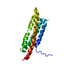 2lk4C 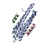 4r32C 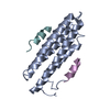 3gm1S C: citing same article ( S: Starting model for refinement |
|---|---|
| Similar structure data |


-Links
 PDBj
PDBj


- Assembly
| Deposited unit | 
| ||||||||
|---|---|---|---|---|---|---|---|---|---|
| 1 | 
| ||||||||
| 2 | 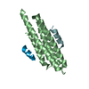
| ||||||||
| 3 | 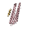
| ||||||||
| 4 | 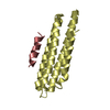
| ||||||||
| Unit cell |
|
-Components
| #1: Protein | Mass: 15260.584 Da / Num. of mol.: 4 / Fragment: unp residues 871-1005 / Mutation: C899S Source method: isolated from a genetically manipulated source Source: (gene. exp.) Homo sapiens (human) / Gene: PTK2B, FAK2, PYK2, RAFTK / Plasmid: pET28 / Production host:  References: UniProt: Q14289, non-specific protein-tyrosine kinase #2: Protein/peptide | Mass: 1915.128 Da / Num. of mol.: 6 / Fragment: unp residues 261-277 / Source method: obtained synthetically / Details: synthetic peptide / Source: (synth.) Homo sapiens (human) / References: UniProt: P49023#3: Water | ChemComp-HOH / |  Mass: 18.015 Da / Num. of mol.: 3 / Source method: isolated from a natural source / Formula: H2O Mass: 18.015 Da / Num. of mol.: 3 / Source method: isolated from a natural source / Formula: H2O |
|---|
-Experimental details
-Experiment
| Experiment | Method: X-RAY DIFFRACTION / Number of used crystals: 1 |
|---|
- Sample preparation
| Crystal | Density Matthews: 4.12 Å3/Da / Density % sol: 70.11 % |
|---|---|
| Crystal grow | Temperature: 291.2 K / Method: vapor diffusion, sitting drop / pH: 6.3 Details: The 4 ul drop contained 2 ul protein-LD4 peptide mixture (20mM Mes, pH6.2, 1mM protein, 2 mM peptide) and 2 ul ML (100 mM MES pH6.3, 4.2 M NaCl, 2%(v/v) glycerol., VAPOR DIFFUSION, SITTING ...Details: The 4 ul drop contained 2 ul protein-LD4 peptide mixture (20mM Mes, pH6.2, 1mM protein, 2 mM peptide) and 2 ul ML (100 mM MES pH6.3, 4.2 M NaCl, 2%(v/v) glycerol., VAPOR DIFFUSION, SITTING DROP, temperature 291.2K |
-Data collection
| Diffraction | Mean temperature: 100 K |
|---|---|
| Diffraction source | Source: SYNCHROTRON / Site: APS  / Beamline: 22-BM / Wavelength: 1 Å / Beamline: 22-BM / Wavelength: 1 Å |
| Detector | Type: MARMOSAIC 225 mm CCD / Detector: CCD / Date: Mar 13, 2011 / Details: mirrors |
| Radiation | Monochromator: Si-220 / Protocol: SINGLE WAVELENGTH / Monochromatic (M) / Laue (L): M / Scattering type: x-ray |
| Radiation wavelength | Wavelength: 1 Å / Relative weight: 1 |
| Reflection | Resolution: 3.1→30 Å / Num. all: 22414 / Num. obs: 21900 / % possible obs: 97.9 % / Observed criterion σ(F): 2 / Observed criterion σ(I): 2 / Redundancy: 5 % / Biso Wilson estimate: 106.2 Å2 / Rsym value: 0.06 / Net I/σ(I): 25.8 |
| Reflection shell | Resolution: 3.1→3.21 Å / Redundancy: 3.9 % / Mean I/σ(I) obs: 1.8 / Num. unique all: 2076 / Rsym value: 0.647 / % possible all: 94.4 |
- Processing
| Software |
| ||||||||||||||||||||||||||||||||||||||||||||||||||||||||
|---|---|---|---|---|---|---|---|---|---|---|---|---|---|---|---|---|---|---|---|---|---|---|---|---|---|---|---|---|---|---|---|---|---|---|---|---|---|---|---|---|---|---|---|---|---|---|---|---|---|---|---|---|---|---|---|---|---|
| Refinement | Method to determine structure: MOLECULAR REPLACEMENT Starting model: PDB Entry 3GM1 Resolution: 3.101→29.848 Å / SU ML: 0.3 / σ(F): 0.08 / Phase error: 29.05 / Stereochemistry target values: ML
| ||||||||||||||||||||||||||||||||||||||||||||||||||||||||
| Solvent computation | Shrinkage radii: 0.9 Å / VDW probe radii: 1.11 Å / Solvent model: FLAT BULK SOLVENT MODEL / Bsol: 78.391 Å2 / ksol: 0.324 e/Å3 | ||||||||||||||||||||||||||||||||||||||||||||||||||||||||
| Displacement parameters |
| ||||||||||||||||||||||||||||||||||||||||||||||||||||||||
| Refinement step | Cycle: LAST / Resolution: 3.101→29.848 Å
| ||||||||||||||||||||||||||||||||||||||||||||||||||||||||
| Refine LS restraints |
| ||||||||||||||||||||||||||||||||||||||||||||||||||||||||
| LS refinement shell |
|
