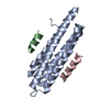
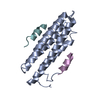
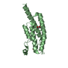
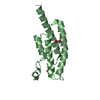
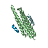
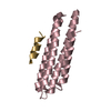
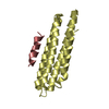
登録情報 データベース : PDB / ID : 3u3fタイトル Structural basis for the interaction of Pyk2 PAT domain with paxillin LD motifs Paxillin LD2 peptide Protein-tyrosine kinase 2-beta キーワード / / / / / 機能・相同性 分子機能 ドメイン・相同性 構成要素
/ / / / / / / / / / / / / / / / / / / / / / / / / / / / / / / / / / / / / / / / / / / / / / / / / / / / / / / / / / / / / / / / / / / / / / / / / / / / / / / / / / / / / / / / / / / / / / / / / / / / / / / / / / / / / / / / / / / / / / / / / / / / / / / / / / / / / / / / / / / / / / / / / / / / / / / / / / / / / / 生物種 Homo sapiens (ヒト)手法 / / / 解像度 : 3.101 Å データ登録者 Vanarotti, M. / Miller, D.J. / Guibao, C.C. / Zheng, J.J. ジャーナル : J.Mol.Biol. / 年 : 2014タイトル : Structural and Mechanistic Insights into the Interaction between Pyk2 and Paxillin LD Motifs.著者 : Vanarotti, M.S. / Miller, D.J. / Guibao, C.D. / Nourse, A. / Zheng, J.J. 履歴 登録 2011年10月5日 登録サイト / 処理サイト 改定 1.0 2012年10月24日 Provider / タイプ 改定 1.1 2014年10月8日 Group 改定 1.2 2014年12月24日 Group 改定 1.3 2023年9月13日 Group / Database references / Refinement descriptionカテゴリ chem_comp_atom / chem_comp_bond ... chem_comp_atom / chem_comp_bond / database_2 / pdbx_initial_refinement_model / struct_ref_seq_dif Item / _database_2.pdbx_database_accession / _struct_ref_seq_dif.details
すべて表示 表示を減らす
 ムービー
ムービー コントローラー


 データを開く
 基本情報
基本情報 要素
要素 キーワード
キーワード focal adhesion /
  機能・相同性情報
 データ登録者
 引用
引用 構造の表示
構造の表示 ダウンロードとリンク
 その他のダウンロード
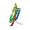


 PDBj
PDBj


 集合体
集合体


 分子量: 18.015 Da / 分子数: 3 / 由来タイプ: 天然 / 式: H2O
 試料調製
試料調製 / ビームライン: 22-BM / 波長: 1 Å
 解析
解析