Mass: 18.015 Da / Num. of mol.: 431 / Source method: isolated from a natural source / Formula: H2O
Has protein modification
Y
Sequence details
THE CONSTRUCT WAS EXPRESSED WITH A PURIFICATION TAG MGSDKIHHHHHHENLYFQG. THE TAG WAS REMOVED WITH ...THE CONSTRUCT WAS EXPRESSED WITH A PURIFICATION TAG MGSDKIHHHHHHENLYFQG. THE TAG WAS REMOVED WITH TEV PROTEASE LEAVING ONLY A GLYCINE (0) FOLLOWED BY RESIDUES 24-258 OF THE TARGET SEQUENCE.
-
Experimental details
-
Experiment
Experiment
Method: X-RAY DIFFRACTION / Number of used crystals: 1
-
Sample preparation
Crystal
Density Matthews: 2 Å3/Da / Density % sol: 38.59 %
Crystal grow
Temperature: 277 K / Method: vapor diffusion, sitting drop Details: 0.1M MES pH 6, 30% polyethylene glycol 6000, NANODROP, VAPOR DIFFUSION, SITTING DROP, temperature 277K
Monochromator: double crystal Si(111) / Protocol: SINGLE WAVELENGTH / Monochromatic (M) / Laue (L): M / Scattering type: x-ray
Radiation wavelength
Wavelength: 0.97907 Å / Relative weight: 1
Reflection
Resolution: 1.7→27.459 Å / Num. obs: 44063 / % possible obs: 93.2 % / Observed criterion σ(I): -3 / Redundancy: 3.9 % / Biso Wilson estimate: 18.403 Å2 / Rmerge(I) obs: 0.051 / Net I/σ(I): 12
Reflection shell
Diffraction-ID: 1
Resolution (Å)
Highest resolution (Å)
Rmerge(I) obs
Mean I/σ(I) obs
Num. measured obs
Num. unique obs
% possible all
1.7-1.76
0.521
1.58
15094
7644
83
1.76-1.83
0.383
2.2
17087
8570
93.7
1.83-1.91
0.268
3
16765
8397
94
1.91-2.02
0.174
4.6
18958
9503
93.7
2.02-2.14
0.119
6.6
16651
8344
94.6
2.14-2.31
0.088
9
17887
8959
94.5
2.31-2.54
0.07
11.2
17284
8666
95
2.54-2.9
0.048
15.6
17147
8593
94.6
2.9-3.66
0.024
26.5
17616
8835
94.3
3.66
0.017
38
17565
8802
94.7
-
Phasing
Phasing
Method: SAD
-
Processing
Software
Name
Version
Classification
NB
MolProbity
3beta29
modelbuilding
PDB_EXTRACT
3.1
dataextraction
SHELX
phasing
SHARP
phasing
XSCALE
December6, 2010
datascaling
REFMAC
5.6.0117
refinement
XDS
datareduction
SHELXD
phasing
autoSHARP
phasing
Refinement
Method to determine structure: SAD / Resolution: 1.7→27.459 Å / Cor.coef. Fo:Fc: 0.967 / Cor.coef. Fo:Fc free: 0.948 / Occupancy max: 1 / Occupancy min: 0.23 / SU B: 4.299 / SU ML: 0.069 / Cross valid method: THROUGHOUT / σ(F): 0 / ESU R Free: 0.108 Stereochemistry target values: MAXIMUM LIKELIHOOD WITH PHASES Details: 1. HYDROGENS HAVE BEEN ADDED IN THE RIDING POSITIONS. 2. A MET-INHIBITION PROTOCOL WAS USED FOR SELENOMETHIONINE INCORPORATION DURING PROTEIN EXPRESSION. THE OCCUPANCY OF THE SE ATOMS IN THE ...Details: 1. HYDROGENS HAVE BEEN ADDED IN THE RIDING POSITIONS. 2. A MET-INHIBITION PROTOCOL WAS USED FOR SELENOMETHIONINE INCORPORATION DURING PROTEIN EXPRESSION. THE OCCUPANCY OF THE SE ATOMS IN THE MSE RESIDUES WAS REDUCED TO 0.75 FOR THE REDUCED SCATTERING POWER DUE TO PARTIAL S-MET INCORPORATION. 3. PEG (PGE) MODELED ARE PRESENT IN CRYO CONDITION. 4. ATOM RECORD CONTAINS SUM OF TLS AND RESIDUAL B FACTORS. ANISOU RECORD CONTAINS SUM OF TLS AND RESIDUAL U FACTORS.
Rfactor
Num. reflection
% reflection
Selection details
Rfree
0.1967
2234
5.1 %
RANDOM
Rwork
0.1578
-
-
-
obs
0.1597
44063
95.18 %
-
Solvent computation
Ion probe radii: 0.8 Å / Shrinkage radii: 0.8 Å / VDW probe radii: 1.2 Å / Solvent model: MASK
In the structure databanks used in Yorodumi, some data are registered as the other names, "COVID-19 virus" and "2019-nCoV". Here are the details of the virus and the list of structure data.
Jan 31, 2019. EMDB accession codes are about to change! (news from PDBe EMDB page)
EMDB accession codes are about to change! (news from PDBe EMDB page)
The allocation of 4 digits for EMDB accession codes will soon come to an end. Whilst these codes will remain in use, new EMDB accession codes will include an additional digit and will expand incrementally as the available range of codes is exhausted. The current 4-digit format prefixed with “EMD-” (i.e. EMD-XXXX) will advance to a 5-digit format (i.e. EMD-XXXXX), and so on. It is currently estimated that the 4-digit codes will be depleted around Spring 2019, at which point the 5-digit format will come into force.
The EM Navigator/Yorodumi systems omit the EMD- prefix.
Related info.:Q: What is EMD? / ID/Accession-code notation in Yorodumi/EM Navigator
Yorodumi is a browser for structure data from EMDB, PDB, SASBDB, etc.
This page is also the successor to EM Navigator detail page, and also detail information page/front-end page for Omokage search.
The word "yorodu" (or yorozu) is an old Japanese word meaning "ten thousand". "mi" (miru) is to see.
Related info.:EMDB / PDB / SASBDB / Comparison of 3 databanks / Yorodumi Search / Aug 31, 2016. New EM Navigator & Yorodumi / Yorodumi Papers / Jmol/JSmol / Function and homology information / Changes in new EM Navigator and Yorodumi
 Movie
Movie Controller

 Yorodumi
 Open data
 Basic information
 Components
 Keywords
 Function and homology information
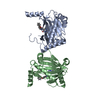
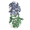
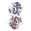
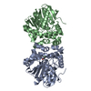
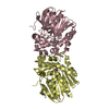
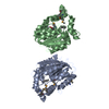
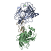
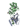
 Parabacteroides distasonis (bacteria)
 X-RAY DIFFRACTION /
  Authors
 Citation
 Structure visualization
 Downloads & links
 Other downloads
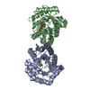
 PDBj
PDBj Assembly
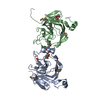

 Mass: 150.173 Da / Num. of mol.: 2 / Source method: obtained synthetically / Formula: C6H14O4
 Mass: 18.015 Da / Num. of mol.: 431 / Source method: isolated from a natural source / Formula: H2O
 Sample preparation
 / Beamline: BL14-1 / Wavelength: 0.97907
  Processing