 Movie
Movie Controller

[English] 日本語
 Yorodumi
- PDB-6qhv: Time resolved structural analysis of the full turnover of an enzy... -
+ Open data

- Basic information
| Entry | Database: PDB / ID: 6qhv | ||||||
|---|---|---|---|---|---|---|---|
| Title | Time resolved structural analysis of the full turnover of an enzyme - 188 ms | ||||||
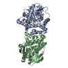
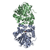
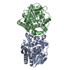
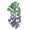
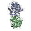
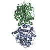
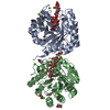
 Components Components | Fluoroacetate dehalogenase | ||||||
 Keywords Keywords | HYDROLASE / time-resolved / catalysis / intermediate | ||||||
| Function / homology |  Function and homology information Function and homology information | ||||||
| Biological species |  Rhodopseudomonas palustris (phototrophic) Rhodopseudomonas palustris (phototrophic) | ||||||
| Method |  X-RAY DIFFRACTION / SYNCHROTRON / MOLECULAR REPLACEMENT / Resolution: 1.715 Å X-RAY DIFFRACTION / SYNCHROTRON / MOLECULAR REPLACEMENT / Resolution: 1.715 Å | ||||||
 Authors Authors | Schulz, E.C. / Mehrabi, P. / Pai, E.F. / Miller, D. | ||||||
 Citation Citation | Journal: Science / Year: 2019 Title: Time-resolved crystallography reveals allosteric communication aligned with molecular breathing. Authors: Mehrabi, P. / Schulz, E.C. / Dsouza, R. / Muller-Werkmeister, H.M. / Tellkamp, F. / Miller, R.J.D. / Pai, E.F. | ||||||
| History |
|
- Structure visualization
| Structure viewer | Molecule: MolmilJmol/JSmol |
|---|
- Downloads & links
-Download
| PDBx/mmCIF format | 6qhv.cif.gz | 140.9 KB | Display | PDBx/mmCIF format |
|---|---|---|---|---|
| PDB format | pdb6qhv.ent.gz | 109.2 KB | Display | PDB format |
| PDBx/mmJSON format | 6qhv.json.gz | Tree view | PDBx/mmJSON format | |
| Others |  Other downloads Other downloads |
-Validation report
| Arichive directory | https://data.pdbj.org/pub/pdb/validation_reports/qh/6qhvftp://data.pdbj.org/pub/pdb/validation_reports/qh/6qhv | HTTPS FTP |
|---|
-Related structure data
| Related structure data | 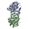 6qhpC 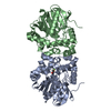 6qhqC 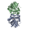 6qhsC 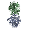 6qhtC 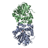 6qhuC 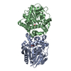 6qhwC 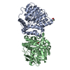 6qhxC  6qhyC 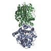 6qhzC 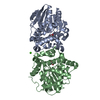 6qi0C 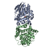 6qi1C 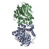 6qi2C  6qi3C 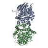 3r3uS S: Starting model for refinement C: citing same article ( |
|---|---|
| Similar structure data |


-Links
 PDBj
PDBj


- Assembly
| Deposited unit | 
| ||||||||
|---|---|---|---|---|---|---|---|---|---|
| 1 |
| ||||||||
| Unit cell |
|
-Components
| #1: Protein | Mass: 34073.660 Da / Num. of mol.: 2 Source method: isolated from a genetically manipulated source Source: (gene. exp.) Rhodopseudomonas palustris (phototrophic)Gene: RPA1163 / Production host: #2: Chemical | ChemComp-FAH / |   Mass: 78.042 Da / Num. of mol.: 1 / Source method: obtained synthetically / Formula: C2H3FO2 / Feature type: SUBJECT OF INVESTIGATION Mass: 78.042 Da / Num. of mol.: 1 / Source method: obtained synthetically / Formula: C2H3FO2 / Feature type: SUBJECT OF INVESTIGATION#3: Water | ChemComp-HOH / |  Mass: 18.015 Da / Num. of mol.: 374 / Source method: isolated from a natural source / Formula: H2O Mass: 18.015 Da / Num. of mol.: 374 / Source method: isolated from a natural source / Formula: H2OHas protein modification | N | |
|---|
-Experimental details
-Experiment
| Experiment | Method: X-RAY DIFFRACTION / Number of used crystals: 1 |
|---|
- Sample preparation
| Crystal | Density Matthews: 1.94 Å3/Da / Density % sol: 36.72 % |
|---|---|
| Crystal grow | Temperature: 293 K / Method: batch mode / pH: 8.5 Details: 18-20 % (w/v)) PEG3350, 200 mM CaCl2, and 100 mM Tris-HCl pH 8.5 |
-Data collection
| Diffraction | Mean temperature: 293 K / Serial crystal experiment: Y |
|---|---|
| Diffraction source | Source: SYNCHROTRON / Site: PETRA III, DESY  / Beamline: P11 / Wavelength: 1.0089 Å / Beamline: P11 / Wavelength: 1.0089 Å |
| Detector | Type: DECTRIS PILATUS 6M / Detector: PIXEL / Date: Apr 15, 2017 |
| Radiation | Protocol: SINGLE WAVELENGTH / Monochromatic (M) / Laue (L): M / Scattering type: x-ray |
| Radiation wavelength | Wavelength: 1.0089 Å / Relative weight: 1 |
| Reflection | Resolution: 1.7→81.5 Å / Num. obs: 53598 / % possible obs: 96.1 % / Redundancy: 94.6 % / CC1/2: 0.974 / Net I/σ(I): 8.1 |
| Reflection shell | Resolution: 1.72→1.82 Å |
| Serial crystallography sample delivery | Description: chip / Method: fixed target |
- Processing
| Software |
| ||||||||||||||||||||||||||||||||||||||||||||||||||||||||||||||||||||||||||||||||||||||||||||||||||||||||||||||||||||||||||||||||||||||||||||
|---|---|---|---|---|---|---|---|---|---|---|---|---|---|---|---|---|---|---|---|---|---|---|---|---|---|---|---|---|---|---|---|---|---|---|---|---|---|---|---|---|---|---|---|---|---|---|---|---|---|---|---|---|---|---|---|---|---|---|---|---|---|---|---|---|---|---|---|---|---|---|---|---|---|---|---|---|---|---|---|---|---|---|---|---|---|---|---|---|---|---|---|---|---|---|---|---|---|---|---|---|---|---|---|---|---|---|---|---|---|---|---|---|---|---|---|---|---|---|---|---|---|---|---|---|---|---|---|---|---|---|---|---|---|---|---|---|---|---|---|---|---|
| Refinement | Method to determine structure: MOLECULAR REPLACEMENT Starting model: 3r3u Resolution: 1.715→19.996 Å / SU ML: 0.17 / Cross valid method: FREE R-VALUE / σ(F): 1.37 / Phase error: 19.59
| ||||||||||||||||||||||||||||||||||||||||||||||||||||||||||||||||||||||||||||||||||||||||||||||||||||||||||||||||||||||||||||||||||||||||||||
| Solvent computation | Shrinkage radii: 0.9 Å / VDW probe radii: 1.11 Å | ||||||||||||||||||||||||||||||||||||||||||||||||||||||||||||||||||||||||||||||||||||||||||||||||||||||||||||||||||||||||||||||||||||||||||||
| Refinement step | Cycle: LAST / Resolution: 1.715→19.996 Å
| ||||||||||||||||||||||||||||||||||||||||||||||||||||||||||||||||||||||||||||||||||||||||||||||||||||||||||||||||||||||||||||||||||||||||||||
| Refine LS restraints |
| ||||||||||||||||||||||||||||||||||||||||||||||||||||||||||||||||||||||||||||||||||||||||||||||||||||||||||||||||||||||||||||||||||||||||||||
| LS refinement shell |
|
