 Movie
Movie Controller

[English] 日本語
 Yorodumi
- PDB-3u0j: Crystal structure of ADP-ribosyltransferase HopU1 of Pseudomonas ... -
+ Open data

- Basic information
| Entry | Database: PDB / ID: 3u0j | ||||||
|---|---|---|---|---|---|---|---|
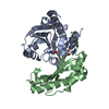
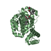
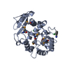
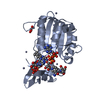
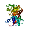
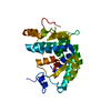
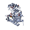
| Title | Crystal structure of ADP-ribosyltransferase HopU1 of Pseudomonas syringae pv. Tomato DC3000 | ||||||
 Components Components | Type III effector HopU1 | ||||||
 Keywords Keywords | TRANSFERASE / ADP-ribosyltransferase / GRP7 | ||||||
| Function / homology |  Function and homology information Function and homology informationNAD+-protein-arginine ADP-ribosyltransferase activity / extracellular region Similarity search - Function | ||||||
| Biological species |  Pseudomonas syringae pv. tomato (bacteria) Pseudomonas syringae pv. tomato (bacteria) | ||||||
| Method |  X-RAY DIFFRACTION / SYNCHROTRON / SAD / Resolution: 2.7 Å X-RAY DIFFRACTION / SYNCHROTRON / SAD / Resolution: 2.7 Å | ||||||
 Authors Authors | Lin, Y. / Yang, H. / Wang, P. / Xu, Y. | ||||||
 Citation Citation | Journal: J.Biol.Chem. / Year: 2011 Title: Structure function analysis of an ADP-ribosyltransferase type III effector and its RNA-binding target in plant immunity Authors: Jeong, B.-R. / Lin, Y. / Joe, A. / Guo, M. / Korneli, C. / Yang, H. / Wang, P. / Yu, M. / Cerny, R.L. / Staiger, D. / Alfano, J.R. / Xu, Y. | ||||||
| History |
|
- Structure visualization
| Structure viewer | Molecule: MolmilJmol/JSmol |
|---|
- Downloads & links
-Download
| PDBx/mmCIF format | 3u0j.cif.gz | 108.5 KB | Display | PDBx/mmCIF format |
|---|---|---|---|---|
| PDB format | pdb3u0j.ent.gz | 83.6 KB | Display | PDB format |
| PDBx/mmJSON format | 3u0j.json.gz | Tree view | PDBx/mmJSON format | |
| Others |  Other downloads Other downloads |
-Validation report
| Arichive directory | https://data.pdbj.org/pub/pdb/validation_reports/u0/3u0jftp://data.pdbj.org/pub/pdb/validation_reports/u0/3u0j | HTTPS FTP |
|---|
-Related structure data
| Similar structure data |
|---|


-Links
 PDBj
PDBj- Assembly
| Deposited unit | 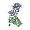
| ||||||||
|---|---|---|---|---|---|---|---|---|---|
| 1 | 
| ||||||||
| 2 | 
| ||||||||
| Unit cell |
|
-Components
| #1: Protein | Mass: 30577.232 Da / Num. of mol.: 2 Source method: isolated from a genetically manipulated source Source: (gene. exp.) Pseudomonas syringae pv. tomato (bacteria)Strain: DC3000 / Gene: hopU1, PSPTO_0501 / Plasmid: pGEX-6P-1 / Production host: #2: Water | ChemComp-HOH / |  Mass: 18.015 Da / Num. of mol.: 205 / Source method: isolated from a natural source / Formula: H2O Mass: 18.015 Da / Num. of mol.: 205 / Source method: isolated from a natural source / Formula: H2O |
|---|
-Experimental details
-Experiment
| Experiment | Method: X-RAY DIFFRACTION / Number of used crystals: 3 |
|---|
- Sample preparation
| Crystal | Density Matthews: 3.56 Å3/Da / Density % sol: 65.44 % |
|---|---|
| Crystal grow | Temperature: 277 K / Method: vapor diffusion, hanging drop / pH: 7.3 Details: 0.1M HEPES (pH 7.3), 5% PEG 10000, 8% Ethylene glycol, VAPOR DIFFUSION, HANGING DROP, temperature 277K |
-Data collection
| Diffraction | Mean temperature: 100 K |
|---|---|
| Diffraction source | Source: SYNCHROTRON / Site: SSRF  / Beamline: BL17U / Wavelength: 0.9788 Å / Beamline: BL17U / Wavelength: 0.9788 Å |
| Detector | Type: MARMOSAIC 225 mm CCD / Detector: CCD / Date: Mar 18, 2010 / Details: mirrors |
| Radiation | Monochromator: SAGITALLY FOCUSED Si(111) / Protocol: SINGLE WAVELENGTH / Monochromatic (M) / Laue (L): M / Scattering type: x-ray |
| Radiation wavelength | Wavelength: 0.9788 Å / Relative weight: 1 |
| Reflection | Resolution: 2.7→50 Å / Num. all: 23703 / Num. obs: 22825 / % possible obs: 96.25 % / Biso Wilson estimate: 26.78 Å2 |
| Reflection shell | Resolution: 2.7→2.8 Å / % possible all: 99.7 |
- Processing
| Software |
| ||||||||||||||||||||||||||||||||||||||||||||||||||||||
|---|---|---|---|---|---|---|---|---|---|---|---|---|---|---|---|---|---|---|---|---|---|---|---|---|---|---|---|---|---|---|---|---|---|---|---|---|---|---|---|---|---|---|---|---|---|---|---|---|---|---|---|---|---|---|---|
| Refinement | Method to determine structure: SAD / Resolution: 2.7→46.279 Å / Occupancy max: 1 / Occupancy min: 1 / SU ML: 0.29 / σ(F): 0.07 / Phase error: 19.46 / Stereochemistry target values: ML
| ||||||||||||||||||||||||||||||||||||||||||||||||||||||
| Solvent computation | Shrinkage radii: 0.9 Å / VDW probe radii: 1.11 Å / Solvent model: FLAT BULK SOLVENT MODEL / Bsol: 19.927 Å2 / ksol: 0.346 e/Å3 | ||||||||||||||||||||||||||||||||||||||||||||||||||||||
| Displacement parameters | Biso max: 110.08 Å2 / Biso mean: 27.4888 Å2 / Biso min: 1.96 Å2
| ||||||||||||||||||||||||||||||||||||||||||||||||||||||
| Refinement step | Cycle: LAST / Resolution: 2.7→46.279 Å
| ||||||||||||||||||||||||||||||||||||||||||||||||||||||
| Refine LS restraints |
| ||||||||||||||||||||||||||||||||||||||||||||||||||||||
| LS refinement shell | Refine-ID: X-RAY DIFFRACTION / Total num. of bins used: 8
|
