 Movie
Movie Controller

[English] 日本語
 Yorodumi
- PDB-3tq5: Crystal structure of M-PMV dUTPASE post-inversion product (dUMP) ... -
+ Open data

- Basic information
| Entry | Database: PDB / ID: 3tq5 | ||||||
|---|---|---|---|---|---|---|---|
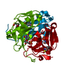
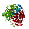
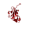
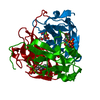
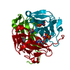
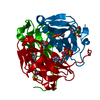
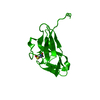
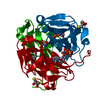
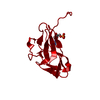
| Title | Crystal structure of M-PMV dUTPASE post-inversion product (dUMP) COMPLEX | ||||||
 Components Components | DEOXYURIDINE 5'-TRIPHOSPHATE NUCLEOTIDO HYDROLASE | ||||||
 Keywords Keywords | HYDROLASE / jelly roll | ||||||
| Function / homology |  Function and homology information Function and homology informationdUTP diphosphatase / dUTP diphosphatase activity / nucleotide metabolic process / Hydrolases; Acting on peptide bonds (peptidases); Aspartic endopeptidases / viral nucleocapsid / structural constituent of virion / aspartic-type endopeptidase activity / viral translational frameshifting / proteolysis / DNA binding / zinc ion binding Similarity search - Function | ||||||
| Biological species |  Mason-Pfizer monkey virus Mason-Pfizer monkey virus | ||||||
| Method |  X-RAY DIFFRACTION / SYNCHROTRON / rigid body refinement / Resolution: 1.4 Å X-RAY DIFFRACTION / SYNCHROTRON / rigid body refinement / Resolution: 1.4 Å | ||||||
 Authors Authors | Barabas, O. / Nemeth, V. / Vertessy, B.G. | ||||||
 Citation Citation | Journal: to be published Title: Structural Snapshots of Enzyme-Catalysed Phosphate Ester Hydrolysis Directly Visualize In-line Attack and Inversion Authors: Barabas, O. / Nemeth, V. / Bodor, A. / Perczel, A. / Rosta, E. / Kele, Z. / Zagyva, I. / Szabadka, Z. / Grolmusz, V.I. / Wilmanns, M. / Vertessy, B.G. #1: Journal: Acta Crystallogr.,Sect.F / Year: 2006 Title: Crystallization and preliminary X-ray studies of dUTPase from Mason-Pfizer monkey retrovirus. Authors: Barabas, O. / Nemeth, V. / Vertessy, B.G. | ||||||
| History |
|
- Structure visualization
| Structure viewer | Molecule: MolmilJmol/JSmol |
|---|
- Downloads & links
-Download
| PDBx/mmCIF format | 3tq5.cif.gz | 65.7 KB | Display | PDBx/mmCIF format |
|---|---|---|---|---|
| PDB format | pdb3tq5.ent.gz | 47.4 KB | Display | PDB format |
| PDBx/mmJSON format | 3tq5.json.gz | Tree view | PDBx/mmJSON format | |
| Others |  Other downloads Other downloads |
-Validation report
| Arichive directory | https://data.pdbj.org/pub/pdb/validation_reports/tq/3tq5ftp://data.pdbj.org/pub/pdb/validation_reports/tq/3tq5 | HTTPS FTP |
|---|
-Related structure data
| Related structure data |  3tp1C  3tpnC  3tpsC  3tpwC 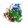 3tpyC 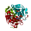 3tq3C  3tq4C 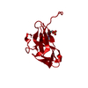 3trlC 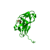 2d4lS S: Starting model for refinement C: citing same article ( |
|---|---|
| Similar structure data |


-Links
 PDBj
PDBj

- Assembly
| Deposited unit | 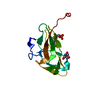
| ||||||||
|---|---|---|---|---|---|---|---|---|---|
| 1 | 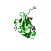
| ||||||||
| Unit cell |
| ||||||||
| Components on special symmetry positions |
|
-Components
| #1: Protein | Mass: 16155.373 Da / Num. of mol.: 1 / Fragment: dUTPase (catalytic) domain, UNP residues 608-759 / Mutation: N1K Source method: isolated from a genetically manipulated source Source: (gene. exp.) Mason-Pfizer monkey virus / Gene: gag-pro / Plasmid: pET22B / Production host:  References: UniProt: O92810, UniProt: P07570*PLUS, dUTP diphosphatase |
|---|---|
| #2: Chemical | ChemComp-UMP /   Mass: 308.182 Da / Num. of mol.: 1 / Source method: obtained synthetically / Formula: C9H13N2O8P Mass: 308.182 Da / Num. of mol.: 1 / Source method: obtained synthetically / Formula: C9H13N2O8P |
| #3: Chemical | ChemComp-TRS /   Mass: 122.143 Da / Num. of mol.: 1 / Source method: obtained synthetically / Formula: C4H12NO3 / Comment: pH buffer*YM Mass: 122.143 Da / Num. of mol.: 1 / Source method: obtained synthetically / Formula: C4H12NO3 / Comment: pH buffer*YM |
| #4: Water | ChemComp-HOH /  Mass: 18.015 Da / Num. of mol.: 107 / Source method: isolated from a natural source / Formula: H2O Mass: 18.015 Da / Num. of mol.: 107 / Source method: isolated from a natural source / Formula: H2O |
-Experimental details
-Experiment
| Experiment | Method: X-RAY DIFFRACTION / Number of used crystals: 1 |
|---|
- Sample preparation
| Crystal | Density Matthews: 2.1 Å3/Da / Density % sol: 41.44 % |
|---|---|
| Crystal grow | Temperature: 293 K / Method: vapor diffusion, hanging drop / pH: 8.5 Details: PEG 8000, AMMONIUM CHLORIDE, TRIS, PH 8.5, vapor diffusion, hanging drop, temperature 293K |
-Data collection
| Diffraction | Mean temperature: 100 K |
|---|---|
| Diffraction source | Source: SYNCHROTRON / Site: EMBL/DESY, HAMBURG  / Beamline: X11 / Wavelength: 0.8128 Å / Beamline: X11 / Wavelength: 0.8128 Å |
| Detector | Type: MAR CCD 165 mm / Detector: CCD / Date: Dec 13, 2004 / Details: mirrors |
| Radiation | Monochromator: Si [111], horizontally focusing / Protocol: SINGLE WAVELENGTH / Monochromatic (M) / Laue (L): M / Scattering type: x-ray |
| Radiation wavelength | Wavelength: 0.8128 Å / Relative weight: 1 |
| Reflection | Resolution: 1.4→20 Å / Num. all: 26148 / Num. obs: 26148 / % possible obs: 99.3 % / Observed criterion σ(I): -3 / Redundancy: 2.7 % / Rsym value: 0.071 / Net I/σ(I): 13.4 |
| Reflection shell | Resolution: 1.4→1.48 Å / Redundancy: 2.2 % / Mean I/σ(I) obs: 2.6 / Num. unique all: 4009 / Rsym value: 0.417 / % possible all: 99.8 |
- Processing
| Software |
| ||||||||||||||||||||||||||||||||||||||||||||||||||||||||||||||||||||||||||||||||||||||||||
|---|---|---|---|---|---|---|---|---|---|---|---|---|---|---|---|---|---|---|---|---|---|---|---|---|---|---|---|---|---|---|---|---|---|---|---|---|---|---|---|---|---|---|---|---|---|---|---|---|---|---|---|---|---|---|---|---|---|---|---|---|---|---|---|---|---|---|---|---|---|---|---|---|---|---|---|---|---|---|---|---|---|---|---|---|---|---|---|---|---|---|---|
| Refinement | Method to determine structure: rigid body refinement Starting model: PDB entry 2D4L Resolution: 1.4→20 Å / Cor.coef. Fo:Fc: 0.973 / Cor.coef. Fo:Fc free: 0.968 / Occupancy max: 1 / Occupancy min: 0.3 / SU B: 3.079 / SU ML: 0.051 / Isotropic thermal model: TLS and isotropic individual / Cross valid method: THROUGHOUT / σ(F): 0 / ESU R Free: 0.056 / Stereochemistry target values: MAXIMUM LIKELIHOOD / Details: HYDROGENS HAVE BEEN ADDED IN THE RIDING POSITIONS
| ||||||||||||||||||||||||||||||||||||||||||||||||||||||||||||||||||||||||||||||||||||||||||
| Solvent computation | Ion probe radii: 0.8 Å / Shrinkage radii: 0.8 Å / VDW probe radii: 1.2 Å / Solvent model: BABINET MODEL WITH MASK | ||||||||||||||||||||||||||||||||||||||||||||||||||||||||||||||||||||||||||||||||||||||||||
| Displacement parameters | Biso max: 59.37 Å2 / Biso mean: 24.752 Å2 / Biso min: 11.98 Å2
| ||||||||||||||||||||||||||||||||||||||||||||||||||||||||||||||||||||||||||||||||||||||||||
| Refinement step | Cycle: LAST / Resolution: 1.4→20 Å
| ||||||||||||||||||||||||||||||||||||||||||||||||||||||||||||||||||||||||||||||||||||||||||
| Refine LS restraints |
| ||||||||||||||||||||||||||||||||||||||||||||||||||||||||||||||||||||||||||||||||||||||||||
| LS refinement shell | Resolution: 1.4→1.437 Å / Total num. of bins used: 20
| ||||||||||||||||||||||||||||||||||||||||||||||||||||||||||||||||||||||||||||||||||||||||||
| Refinement TLS params. | Method: refined / Origin x: 2.466 Å / Origin y: 58.6536 Å / Origin z: 30.1581 Å
| ||||||||||||||||||||||||||||||||||||||||||||||||||||||||||||||||||||||||||||||||||||||||||
| Refinement TLS group |
|
