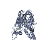
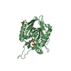
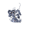
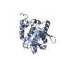
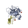
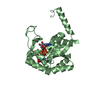
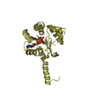
Factors involved in megakaryocyte development and platelet production / Interconversion of nucleotide di- and triphosphates / adenylate kinase / AMP kinase activity / generation of precursor metabolites and energy / mitochondrion / ATP binding / cytosol / cytoplasm Similarity search - Function
Single alpha-helices involved in coiled-coils or other helix-helix interfaces - #2370 / Adenylate kinase, active site lid domain / Adenylate kinase, active site lid / Adenylate kinase subfamily / Adenylate kinase, conserved site / Adenylate kinase signature. / Adenylate kinase/UMP-CMP kinase / Adenylate kinase / Single alpha-helices involved in coiled-coils or other helix-helix interfaces / Helix non-globular ...Single alpha-helices involved in coiled-coils or other helix-helix interfaces - #2370 / Adenylate kinase, active site lid domain / Adenylate kinase, active site lid / Adenylate kinase subfamily / Adenylate kinase, conserved site / Adenylate kinase signature. / Adenylate kinase/UMP-CMP kinase / Adenylate kinase / Single alpha-helices involved in coiled-coils or other helix-helix interfaces / Helix non-globular / Special / P-loop containing nucleotide triphosphate hydrolases / Rossmann fold / P-loop containing nucleoside triphosphate hydrolase / 3-Layer(aba) Sandwich / Alpha Beta Similarity search - Domain/homology
Method: X-RAY DIFFRACTION / Number of used crystals: 1
-
Sample preparation
Crystal
Density Matthews: 2.59 Å3/Da / Density % sol: 52.56 %
Crystal grow
Temperature: 293 K / Method: vapor diffusion, sitting drop / pH: 7.5 Details: 25% PEG 3350, 0.2 M Ammonium Acetate, 0.1 M Hepes, 5 mM ADP, 5 mM MgCl2, 0.3 mM glyglyglycine, VAPOR DIFFUSION, SITTING DROP, temperature 293K, pH 7.5
Method to determine structure: MOLECULAR REPLACEMENT / Resolution: 2.75→34.56 Å / Cor.coef. Fo:Fc: 0.9204 / Cor.coef. Fo:Fc free: 0.8894 / Occupancy max: 1 / Occupancy min: 0.3 / Cross valid method: THROUGHOUT / σ(F): 0 / Stereochemistry target values: Engh & Huber Details: MOLECULAR REPLACEMENT WAS SUCCESSFUL IN FINDING TWO OF FOUR MOLECULES IN THE ASYMMETRIC UNIT, WHICH REFINE WELL AND HAD INTERPRETABLE ELECTRON DENSITY THAT MATCHED THE QUALITY AND RESOLUTION ...Details: MOLECULAR REPLACEMENT WAS SUCCESSFUL IN FINDING TWO OF FOUR MOLECULES IN THE ASYMMETRIC UNIT, WHICH REFINE WELL AND HAD INTERPRETABLE ELECTRON DENSITY THAT MATCHED THE QUALITY AND RESOLUTION OF THE DATA. TWO ADDITIONAL MOLECULES WERE FOUND BY HAND BUILDING INTO SEVERAL AREAS OF ADDITIONAL POSITIVE DENSITY AND TRANSFORMING PARTS OF MOLECULE A ONTO THE BUILT PARTIAL MODEL. NCS AND STRICT GEOMETRIC RESTRAINTS WERE USED THROUGHOUT REFINEMENT AFTER THIS. THE AUTHORS BELIEVE THAT THE POOR DENSITY FOR C AND D ARE DUE TO MARGINAL CRYSTAL PACKING.
Rfactor
Num. reflection
% reflection
Selection details
Rfree
0.2596
1503
5.07 %
RANDOM
Rwork
0.2255
-
-
-
obs
0.2272
29652
-
-
all
-
29681
-
-
Displacement parameters
Biso mean: 100.31 Å2
Baniso -1
Baniso -2
Baniso -3
1-
15.8282 Å2
0 Å2
-2.4215 Å2
2-
-
7.021 Å2
0 Å2
3-
-
-
-22.8492 Å2
Refine analyze
Luzzati coordinate error obs: 0.521 Å
Refinement step
Cycle: LAST / Resolution: 2.75→34.56 Å
Protein
Nucleic acid
Ligand
Solvent
Total
Num. atoms
5630
0
137
0
5767
Refine LS restraints
Refine-ID
Type
Dev ideal
Number
Restraint function
Weight
X-RAY DIFFRACTION
t_bond_d
0.01
5916
HARMONIC
2
X-RAY DIFFRACTION
t_angle_deg
1.18
8099
HARMONIC
2
X-RAY DIFFRACTION
t_dihedral_angle_d
1778
SINUSOIDAL
2
X-RAY DIFFRACTION
t_incorr_chiral_ct
X-RAY DIFFRACTION
t_pseud_angle
X-RAY DIFFRACTION
t_trig_c_planes
123
HARMONIC
2
X-RAY DIFFRACTION
t_gen_planes
897
HARMONIC
5
X-RAY DIFFRACTION
t_it
5742
HARMONIC
20
X-RAY DIFFRACTION
t_nbd
0
SEMIHARMONIC
5
X-RAY DIFFRACTION
t_omega_torsion
2.47
X-RAY DIFFRACTION
t_other_torsion
22.38
X-RAY DIFFRACTION
t_improper_torsion
24
HARMONIC
0
X-RAY DIFFRACTION
t_chiral_improper_torsion
826
SEMIHARMONIC
5
X-RAY DIFFRACTION
t_sum_occupancies
X-RAY DIFFRACTION
t_utility_distance
X-RAY DIFFRACTION
t_utility_angle
X-RAY DIFFRACTION
t_utility_torsion
X-RAY DIFFRACTION
t_ideal_dist_contact
6540
SEMIHARMONIC
4
LS refinement shell
Resolution: 2.75→2.85 Å / Total num. of bins used: 15
Rfactor
Num. reflection
% reflection
Rfree
0.2788
150
5.22 %
Rwork
0.2345
2725
-
all
0.2367
2875
-
Refinement TLS params.
Method: refined / Refine-ID: X-RAY DIFFRACTION
ID
L11 (°2)
L12 (°2)
L13 (°2)
L22 (°2)
L23 (°2)
L33 (°2)
S11 (Å °)
S12 (Å °)
S13 (Å °)
S21 (Å °)
S22 (Å °)
S23 (Å °)
S31 (Å °)
S32 (Å °)
S33 (Å °)
T11 (Å2)
T12 (Å2)
T13 (Å2)
T22 (Å2)
T23 (Å2)
T33 (Å2)
Origin x (Å)
Origin y (Å)
Origin z (Å)
1
6.6144
0.7404
-0.3079
3.4554
-0.6314
2.1249
0.0672
0.4276
-0.4602
-0.2978
-0.1186
0.1784
0.105
-0.1582
0.0513
-0.3262
0.0071
0.0501
-0.2675
0.0091
0.0927
0.362
-13.7617
-2.5329
2
3.71
-2.0605
-0.2223
6.2253
1.0752
1.6646
0.1794
-0.0683
0.1684
0.0584
-0.058
-0.7017
0.0449
-0.0864
-0.1213
-0.1902
-0.0113
0.0117
-0.3164
0.0539
0.1524
37.7743
-2.4312
2.6111
3
3.5882
-0.8499
-1.7065
0
1.4233
10.0322
-0.2335
-0.2901
0.4316
-0.2055
-0.2891
0.0101
0.0163
-0.1936
0.5226
0.0808
0.1515
0.1094
-0.2718
0.0907
-0.1837
1.2714
8.3414
52.0197
4
1.7743
0.0037
-1.7837
0
-0.818
13.1627
0.0085
-0.4136
-0.036
-0.3341
0.31
0.3045
0.4003
1.0293
-0.3185
0.2655
0.1854
-0.0612
0.1025
-0.0573
-0.4155
22.5952
-14.0612
56.4835
Refinement TLS group
ID
Refine-ID
Refine TLS-ID
Selection details
Auth asym-ID
Auth seq-ID
1
X-RAY DIFFRACTION
1
{ A|* }
A
8 - 242
2
X-RAY DIFFRACTION
2
{ B|* }
B
3 - 242
3
X-RAY DIFFRACTION
3
{ C|* }
C
3 - 241
4
X-RAY DIFFRACTION
4
{ D|* }
D
6 - 242
+
About Yorodumi
-
News
-
Feb 9, 2022. New format data for meta-information of EMDB entries
New format data for meta-information of EMDB entries
Version 3 of the EMDB header file is now the official format.
The previous official version 1.9 will be removed from the archive.
In the structure databanks used in Yorodumi, some data are registered as the other names, "COVID-19 virus" and "2019-nCoV". Here are the details of the virus and the list of structure data.
Jan 31, 2019. EMDB accession codes are about to change! (news from PDBe EMDB page)
EMDB accession codes are about to change! (news from PDBe EMDB page)
The allocation of 4 digits for EMDB accession codes will soon come to an end. Whilst these codes will remain in use, new EMDB accession codes will include an additional digit and will expand incrementally as the available range of codes is exhausted. The current 4-digit format prefixed with “EMD-” (i.e. EMD-XXXX) will advance to a 5-digit format (i.e. EMD-XXXXX), and so on. It is currently estimated that the 4-digit codes will be depleted around Spring 2019, at which point the 5-digit format will come into force.
The EM Navigator/Yorodumi systems omit the EMD- prefix.
Related info.:Q: What is EMD? / ID/Accession-code notation in Yorodumi/EM Navigator
Yorodumi is a browser for structure data from EMDB, PDB, SASBDB, etc.
This page is also the successor to EM Navigator detail page, and also detail information page/front-end page for Omokage search.
The word "yorodu" (or yorozu) is an old Japanese word meaning "ten thousand". "mi" (miru) is to see.
Related info.:EMDB / PDB / SASBDB / Comparison of 3 databanks / Yorodumi Search / Aug 31, 2016. New EM Navigator & Yorodumi / Yorodumi Papers / Jmol/JSmol / Function and homology information / Changes in new EM Navigator and Yorodumi
 Movie
Movie Controller

 Yorodumi
 Open data
 Basic information
 Components
 Keywords
 Function and homology information
 X-RAY DIFFRACTION /
  Authors
 Citation
 Structure visualization
 Downloads & links
 Other downloads


 PDBj
PDBj


 Assembly
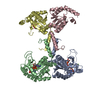


 Mass: 24.305 Da / Num. of mol.: 2 / Source method: obtained synthetically / Formula: Mg
 Mass: 347.221 Da / Num. of mol.: 1 / Source method: obtained synthetically / Formula: C10H14N5O7P / Comment: AMP*YM
 Mass: 427.201 Da / Num. of mol.: 3 / Source method: obtained synthetically / Formula: C10H15N5O10P2 / Comment: ADP, energy-carrying molecule*YM
 Mass: 507.181 Da / Num. of mol.: 1 / Source method: obtained synthetically / Formula: C10H16N5O13P3 / Comment: ATP, energy-carrying molecule*YM
 Sample preparation
 / Beamline: 19-ID / Wavelength: 0.97911 Å
 Processing