 Movie
Movie Controller

[English] 日本語
 Yorodumi
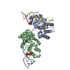
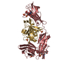
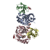
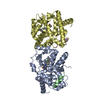
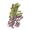
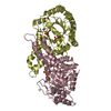
- PDB-3tjz: Crystal Structure of Arf1 Bound to the gamma/zeta-COP Core Complex -
+ Open data

- Basic information
| Entry | Database: PDB / ID: 3tjz | ||||||
|---|---|---|---|---|---|---|---|
| Title | Crystal Structure of Arf1 Bound to the gamma/zeta-COP Core Complex | ||||||
 Components Components |
| ||||||
 Keywords Keywords | protein transport/protein binding / protein trafficking / Golgi membrane / protein transport-protein binding complex | ||||||
| Function / homology |  Function and homology information Function and homology informationSynthesis of PIPs at the plasma membrane / VxPx cargo-targeting to cilium / Synthesis of PIPs at the Golgi membrane / protein localization to Golgi membrane / COPI-dependent Golgi-to-ER retrograde traffic / regulation of Golgi organization / COPI-mediated anterograde transport / trans-Golgi Network Vesicle Budding / organelle membrane contact site / Intra-Golgi traffic ...Synthesis of PIPs at the plasma membrane / VxPx cargo-targeting to cilium / Synthesis of PIPs at the Golgi membrane / protein localization to Golgi membrane / COPI-dependent Golgi-to-ER retrograde traffic / regulation of Golgi organization / COPI-mediated anterograde transport / trans-Golgi Network Vesicle Budding / organelle membrane contact site / Intra-Golgi traffic / COPI vesicle coat / Golgi vesicle transport / COPI-dependent Golgi-to-ER retrograde traffic / COPI-mediated anterograde transport / positive regulation of mitochondrial fusion / organelle transport along microtubule / regulation of fatty acid metabolic process / Golgi to plasma membrane transport / intra-Golgi vesicle-mediated transport / retrograde vesicle-mediated transport, Golgi to endoplasmic reticulum / positive regulation of mitochondrial fission / endoplasmic reticulum-Golgi intermediate compartment / protein secretion / endoplasmic reticulum to Golgi vesicle-mediated transport / vesicle-mediated transport / small monomeric GTPase / macroautophagy / intracellular protein transport / G protein activity / Golgi membrane / GTPase activity / GTP binding / structural molecule activity / endoplasmic reticulum / Golgi apparatus / plasma membrane / cytoplasm / cytosol Similarity search - Function | ||||||
| Biological species |   | ||||||
| Method |  X-RAY DIFFRACTION / SYNCHROTRON / SAD / Resolution: 2.9 Å X-RAY DIFFRACTION / SYNCHROTRON / SAD / Resolution: 2.9 Å | ||||||
 Authors Authors | Goldberg, J. / Yu, X. / Breitman, M. | ||||||
 Citation Citation | Journal: Cell(Cambridge,Mass.) / Year: 2012 Title: A structure-based mechanism for arf1-dependent recruitment of coatomer to membranes. Authors: Yu, X. / Breitman, M. / Goldberg, J. | ||||||
| History |
|
- Structure visualization
| Structure viewer | Molecule: MolmilJmol/JSmol |
|---|
- Downloads & links
-Download
| PDBx/mmCIF format | 3tjz.cif.gz | 235.7 KB | Display | PDBx/mmCIF format |
|---|---|---|---|---|
| PDB format | pdb3tjz.ent.gz | 187.7 KB | Display | PDB format |
| PDBx/mmJSON format | 3tjz.json.gz | Tree view | PDBx/mmJSON format | |
| Others |  Other downloads Other downloads |
-Validation report
| Arichive directory | https://data.pdbj.org/pub/pdb/validation_reports/tj/3tjzftp://data.pdbj.org/pub/pdb/validation_reports/tj/3tjz | HTTPS FTP |
|---|
-Related structure data
| Similar structure data |
|---|


-Links
 PDBj
PDBj


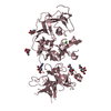
- Assembly
| Deposited unit | 
| ||||||||
|---|---|---|---|---|---|---|---|---|---|
| 1 | 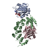
| ||||||||
| 2 | 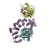
| ||||||||
| Unit cell |
|
-Components
| #1: Protein | Mass: 18665.225 Da / Num. of mol.: 2 / Fragment: unp residues 18-181 Source method: isolated from a genetically manipulated source Source: (gene. exp.) Strain: ATCC 204508 / S288c / Gene: ARF1, YDL192W, D1244 / Plasmid: pET28b / Production host:  #2: Protein | Mass: 39375.480 Da / Num. of mol.: 2 / Fragment: unp residues 1-355 Source method: isolated from a genetically manipulated source Source: (gene. exp.)   Spodoptera frugiperda (fall armyworm) / References: UniProt: P53620 Spodoptera frugiperda (fall armyworm) / References: UniProt: P53620#3: Protein | Mass: 17479.992 Da / Num. of mol.: 2 / Fragment: unp residues 1-153 Source method: isolated from a genetically manipulated source Source: (gene. exp.) Spodoptera frugiperda (fall armyworm) / References: UniProt: P35604#4: Chemical |   Mass: 522.196 Da / Num. of mol.: 2 / Source method: obtained synthetically / Formula: C10H17N6O13P3 Mass: 522.196 Da / Num. of mol.: 2 / Source method: obtained synthetically / Formula: C10H17N6O13P3Comment: GppNHp, GMPPNP, energy-carrying molecule analogue*YM #5: Chemical |   Mass: 24.305 Da / Num. of mol.: 2 / Source method: obtained synthetically / Formula: Mg Mass: 24.305 Da / Num. of mol.: 2 / Source method: obtained synthetically / Formula: Mg |
|---|
-Experimental details
-Experiment
| Experiment | Method: X-RAY DIFFRACTION / Number of used crystals: 1 |
|---|
- Sample preparation
| Crystal | Density Matthews: 3.22 Å3/Da / Density % sol: 61.74 % |
|---|---|
| Crystal grow | Temperature: 298 K / Method: vapor diffusion, hanging drop / pH: 6.5 Details: 9% PEG 400, 0.1 M MES-NaOH pH 6.5, VAPOR DIFFUSION, HANGING DROP, temperature 298K |
-Data collection
| Diffraction | Mean temperature: 100 K |
|---|---|
| Diffraction source | Source: SYNCHROTRON / Site: NSLS  / Beamline: X29A / Wavelength: 1.075 Å / Beamline: X29A / Wavelength: 1.075 Å |
| Detector | Type: ADSC QUANTUM 315r / Detector: CCD / Date: Jun 2, 2010 |
| Radiation | Protocol: SINGLE WAVELENGTH / Monochromatic (M) / Laue (L): M / Scattering type: x-ray |
| Radiation wavelength | Wavelength: 1.075 Å / Relative weight: 1 |
| Reflection | Resolution: 2.9→45 Å / Num. all: 83327 / Num. obs: 83327 / % possible obs: 99.9 % / Redundancy: 7.1 % / Rmerge(I) obs: 0.061 / Net I/σ(I): 32.1 |
| Reflection shell | Resolution: 2.9→3 Å / Redundancy: 7.1 % / Rmerge(I) obs: 0.432 / Mean I/σ(I) obs: 4.8 / % possible all: 100 |
- Processing
| Software |
| ||||||||||||||||||||||||||||||||||||||||||||||||||||||||||||||||||||||||||||||||||||||||||||||||||||||||||||||||||||||||||||||||||||||||||||||||||||||||||||||||||||||||||||||||||||||||||||||||||||
|---|---|---|---|---|---|---|---|---|---|---|---|---|---|---|---|---|---|---|---|---|---|---|---|---|---|---|---|---|---|---|---|---|---|---|---|---|---|---|---|---|---|---|---|---|---|---|---|---|---|---|---|---|---|---|---|---|---|---|---|---|---|---|---|---|---|---|---|---|---|---|---|---|---|---|---|---|---|---|---|---|---|---|---|---|---|---|---|---|---|---|---|---|---|---|---|---|---|---|---|---|---|---|---|---|---|---|---|---|---|---|---|---|---|---|---|---|---|---|---|---|---|---|---|---|---|---|---|---|---|---|---|---|---|---|---|---|---|---|---|---|---|---|---|---|---|---|---|---|---|---|---|---|---|---|---|---|---|---|---|---|---|---|---|---|---|---|---|---|---|---|---|---|---|---|---|---|---|---|---|---|---|---|---|---|---|---|---|---|---|---|---|---|---|---|---|---|---|
| Refinement | Method to determine structure: SAD / Resolution: 2.9→45 Å / SU ML: 0.85 / σ(F): 1.34 / Phase error: 26.22 / Stereochemistry target values: ML
| ||||||||||||||||||||||||||||||||||||||||||||||||||||||||||||||||||||||||||||||||||||||||||||||||||||||||||||||||||||||||||||||||||||||||||||||||||||||||||||||||||||||||||||||||||||||||||||||||||||
| Solvent computation | Shrinkage radii: 0.72 Å / VDW probe radii: 1 Å / Solvent model: FLAT BULK SOLVENT MODEL / Bsol: 54.642 Å2 / ksol: 0.319 e/Å3 | ||||||||||||||||||||||||||||||||||||||||||||||||||||||||||||||||||||||||||||||||||||||||||||||||||||||||||||||||||||||||||||||||||||||||||||||||||||||||||||||||||||||||||||||||||||||||||||||||||||
| Displacement parameters |
| ||||||||||||||||||||||||||||||||||||||||||||||||||||||||||||||||||||||||||||||||||||||||||||||||||||||||||||||||||||||||||||||||||||||||||||||||||||||||||||||||||||||||||||||||||||||||||||||||||||
| Refinement step | Cycle: LAST / Resolution: 2.9→45 Å
| ||||||||||||||||||||||||||||||||||||||||||||||||||||||||||||||||||||||||||||||||||||||||||||||||||||||||||||||||||||||||||||||||||||||||||||||||||||||||||||||||||||||||||||||||||||||||||||||||||||
| Refine LS restraints |
| ||||||||||||||||||||||||||||||||||||||||||||||||||||||||||||||||||||||||||||||||||||||||||||||||||||||||||||||||||||||||||||||||||||||||||||||||||||||||||||||||||||||||||||||||||||||||||||||||||||
| LS refinement shell |
|
