 Movie
Movie Controller

[English] 日本語
 Yorodumi
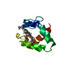
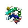
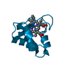
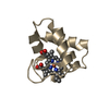
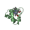
- PDB-3tjy: Structure of the Pto-binding domain of HopPmaL generated by limit... -
+ Open data

- Basic information
| Entry | Database: PDB / ID: 3tjy | ||||||
|---|---|---|---|---|---|---|---|
| Title | Structure of the Pto-binding domain of HopPmaL generated by limited chymotrypsin digestion | ||||||
 Components Components | Effector protein hopAB3 | ||||||
 Keywords Keywords | SIGNALING PROTEIN / type III effector / HopPmaL / Pseudomonas syringae / Structural Genomics / PSI-Biology / Midwest Center for Structural Genomics / MCSG / helical bundle / Pto | ||||||
| Function / homology |  Function and homology information Function and homology informationsymbiont-mediated perturbation of host programmed cell death / extracellular region Similarity search - Function | ||||||
| Biological species |  Pseudomonas syringae pv. maculicola (bacteria) Pseudomonas syringae pv. maculicola (bacteria) | ||||||
| Method |  X-RAY DIFFRACTION / SYNCHROTRON / SAD / Resolution: 1.7 Å X-RAY DIFFRACTION / SYNCHROTRON / SAD / Resolution: 1.7 Å | ||||||
 Authors Authors | Singer, A.U. / Stein, A. / Xu, X. / Cui, H. / Joachimiak, A. / Edwards, A.M. / Savchenko, A. / Midwest Center for Structural Genomics (MCSG) | ||||||
 Citation Citation | Journal: Biochemistry / Year: 2012 Title: Structural analysis of HopPmaL reveals the presence of a second adaptor domain common to the HopAB family of Pseudomonas syringae type III effectors. Authors: Singer, A.U. / Wu, B. / Yee, A. / Houliston, S. / Xu, X. / Cui, H. / Skarina, T. / Garcia, M. / Semesi, A. / Arrowsmith, C.H. / Savchenko, A. | ||||||
| History |
|
- Structure visualization
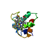
| Structure viewer | Molecule: MolmilJmol/JSmol |
|---|
- Downloads & links
-Download
| PDBx/mmCIF format | 3tjy.cif.gz | 49 KB | Display | PDBx/mmCIF format |
|---|---|---|---|---|
| PDB format | pdb3tjy.ent.gz | 35.9 KB | Display | PDB format |
| PDBx/mmJSON format | 3tjy.json.gz | Tree view | PDBx/mmJSON format | |
| Others |  Other downloads Other downloads |
-Validation report
| Arichive directory | https://data.pdbj.org/pub/pdb/validation_reports/tj/3tjyftp://data.pdbj.org/pub/pdb/validation_reports/tj/3tjy | HTTPS FTP |
|---|
-Related structure data
| Related structure data | 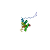 2lf3C 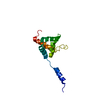 2lf6C 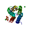 3sviC C: citing same article ( |
|---|---|
| Similar structure data | |
| Other databases |


-Links
 PDBj
PDBj- Assembly
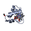
| Deposited unit | 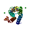
| ||||||||
|---|---|---|---|---|---|---|---|---|---|
| 1 |
| ||||||||
| Unit cell |
|
-Components
| #1: Protein | Mass: 10655.572 Da / Num. of mol.: 1 / Fragment: HopPmaL / Mutation: none Source method: isolated from a genetically manipulated source Source: (gene. exp.) Pseudomonas syringae pv. maculicola (bacteria)Strain: ES4326 / Gene: hopAB3, hopPmaL / Plasmid: p15TvLic / Production host: | ||||||||
|---|---|---|---|---|---|---|---|---|---|
| #2: Chemical |   Mass: 96.063 Da / Num. of mol.: 3 / Source method: obtained synthetically / Formula: SO4 Mass: 96.063 Da / Num. of mol.: 3 / Source method: obtained synthetically / Formula: SO4#3: Chemical | ChemComp-CL /   Mass: 35.453 Da / Num. of mol.: 4 / Source method: obtained synthetically / Formula: Cl Mass: 35.453 Da / Num. of mol.: 4 / Source method: obtained synthetically / Formula: Cl#4: Water | ChemComp-HOH / |  Mass: 18.015 Da / Num. of mol.: 80 / Source method: isolated from a natural source / Formula: H2O Mass: 18.015 Da / Num. of mol.: 80 / Source method: isolated from a natural source / Formula: H2OHas protein modification | Y | Sequence details | PROTEIN WAS CLONED AS HOPPMAL RESIDUES 135-273, CUT WITH TEV, THEN TREATED WITH LIMITING AMOUNTS OF ...PROTEIN WAS CLONED AS HOPPMAL RESIDUES 135-273, CUT WITH TEV, THEN TREATED WITH LIMITING AMOUNTS OF THERMOLYSI | |
-Experimental details
-Experiment
| Experiment | Method: X-RAY DIFFRACTION / Number of used crystals: 1 |
|---|
- Sample preparation
| Crystal | Density Matthews: 2.12 Å3/Da / Density % sol: 41.98 % |
|---|---|
| Crystal grow | Temperature: 294 K / Method: vapor diffusion, hanging drop / pH: 6.5 Details: 0.1M Bis-Tris pH 6.5 plus 1.5M Ammonium Sulphate. 0.03 mg/ml chymotrypsin was added to the protein prior to adding crystallization liquor. Crystals were cryoprotected with Paratone-N oil , ...Details: 0.1M Bis-Tris pH 6.5 plus 1.5M Ammonium Sulphate. 0.03 mg/ml chymotrypsin was added to the protein prior to adding crystallization liquor. Crystals were cryoprotected with Paratone-N oil , VAPOR DIFFUSION, HANGING DROP, temperature 294K |
-Data collection
| Diffraction | Mean temperature: 100 K |
|---|---|
| Diffraction source | Source: SYNCHROTRON / Site: APS  / Beamline: 19-ID / Wavelength: 0.97942 Å / Beamline: 19-ID / Wavelength: 0.97942 Å |
| Detector | Type: ADSC QUANTUM 315 / Detector: CCD / Date: Jul 10, 2009 / Details: mirrors |
| Radiation | Monochromator: SI-111 CHANNEL / Protocol: SINGLE WAVELENGTH / Monochromatic (M) / Laue (L): M / Scattering type: x-ray |
| Radiation wavelength | Wavelength: 0.97942 Å / Relative weight: 1 |
| Reflection | Resolution: 1.65→25.682 Å / Num. all: 11562 / Num. obs: 11555 / % possible obs: 99.9 % / Observed criterion σ(I): -3 / Redundancy: 9.3 % / Rmerge(I) obs: 0.08 / Rsym value: 0.08 / Net I/σ(I): 42.133 |
| Reflection shell | Resolution: 1.65→1.68 Å / Redundancy: 9.5 % / Rmerge(I) obs: 0.501 / Mean I/σ(I) obs: 3.7 / Rsym value: 0.501 / % possible all: 100 |
- Processing
| Software | Name: PHENIX / Version: (phenix.refine: 1.7.1_743) / Classification: refinement | ||||||||||||||||||||||||||||||||||||||||
|---|---|---|---|---|---|---|---|---|---|---|---|---|---|---|---|---|---|---|---|---|---|---|---|---|---|---|---|---|---|---|---|---|---|---|---|---|---|---|---|---|---|
| Refinement | Method to determine structure: SAD / Resolution: 1.7→25.682 Å / SU ML: 0.42 / σ(F): 1.35 / Phase error: 17.62 / Stereochemistry target values: ML
| ||||||||||||||||||||||||||||||||||||||||
| Solvent computation | Shrinkage radii: 0.95 Å / VDW probe radii: 1.2 Å / Solvent model: FLAT BULK SOLVENT MODEL / Bsol: 41.325 Å2 / ksol: 0.39 e/Å3 | ||||||||||||||||||||||||||||||||||||||||
| Displacement parameters |
| ||||||||||||||||||||||||||||||||||||||||
| Refinement step | Cycle: LAST / Resolution: 1.7→25.682 Å
| ||||||||||||||||||||||||||||||||||||||||
| Refine LS restraints |
| ||||||||||||||||||||||||||||||||||||||||
| LS refinement shell |
| ||||||||||||||||||||||||||||||||||||||||
| Refinement TLS params. | Method: refined / Origin x: -14.4761 Å / Origin y: -4.9353 Å / Origin z: 13.9018 Å
| ||||||||||||||||||||||||||||||||||||||||
| Refinement TLS group | Selection details: (chain A and resid 140:217) |
