 Movie
Movie Controller

[English] 日本語
 Yorodumi
- PDB-3tj1: Crystal Structure of RNA Polymerase I Transcription Initiation Fa... -
+ Open data

- Basic information
| Entry | Database: PDB / ID: 3tj1 | ||||||
|---|---|---|---|---|---|---|---|
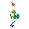
| Title | Crystal Structure of RNA Polymerase I Transcription Initiation Factor Rrn3 | ||||||
 Components Components | RNA polymerase I-specific transcription initiation factor RRN3 | ||||||
 Keywords Keywords | TRANSCRIPTION / HEAT repeat / Transcription Factor / Nucleus | ||||||
| Function / homology |  Function and homology information Function and homology informationRNA polymerase I general transcription initiation factor activity / RNA polymerase I core binding / RNA polymerase I general transcription initiation factor binding / rDNA binding / RNA Polymerase I Transcription Initiation / RNA Polymerase I Promoter Escape / transcription initiation at RNA polymerase I promoter / nucleolus / nucleus Similarity search - Function | ||||||
| Biological species |  | ||||||
| Method |  X-RAY DIFFRACTION / SYNCHROTRON / MAD / Resolution: 2.85 Å X-RAY DIFFRACTION / SYNCHROTRON / MAD / Resolution: 2.85 Å | ||||||
 Authors Authors | Blattner, C. / Jennebach, S. / Herzog, F. / Mayer, A. / Cheung, A.C.M. / Witte, G. / Lorenzen, K. / Hopfner, K.-P. / Heck, A.J.R. / Aebersold, R. / Cramer, P. | ||||||
 Citation Citation | Journal: Genes Dev. / Year: 2011 Title: Molecular basis of Rrn3-regulated RNA polymerase I initiation and cell growth. Authors: Blattner, C. / Jennebach, S. / Herzog, F. / Mayer, A. / Cheung, A.C. / Witte, G. / Lorenzen, K. / Hopfner, K.P. / Heck, A.J. / Aebersold, R. / Cramer, P. | ||||||
| History |
|
- Structure visualization
| Structure viewer | Molecule: MolmilJmol/JSmol |
|---|
- Downloads & links
-Download
| PDBx/mmCIF format | 3tj1.cif.gz | 409 KB | Display | PDBx/mmCIF format |
|---|---|---|---|---|
| PDB format | pdb3tj1.ent.gz | 338.2 KB | Display | PDB format |
| PDBx/mmJSON format | 3tj1.json.gz | Tree view | PDBx/mmJSON format | |
| Others |  Other downloads Other downloads |
-Validation report
| Arichive directory | https://data.pdbj.org/pub/pdb/validation_reports/tj/3tj1ftp://data.pdbj.org/pub/pdb/validation_reports/tj/3tj1 | HTTPS FTP |
|---|
-Related structure data
| Similar structure data |
|---|


-Links
 PDBj
PDBj


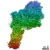
- Assembly
| Deposited unit | 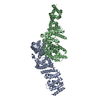
| ||||||||
|---|---|---|---|---|---|---|---|---|---|
| 1 |
| ||||||||
| Unit cell |
|
-Components
| #1: Protein | Mass: 74831.883 Da / Num. of mol.: 2 Source method: isolated from a genetically manipulated source Source: (gene. exp.) Strain: ATCC 204508 / S288c / Gene: RRN3, YKL125W / Plasmid: pET28 / Production host:  #2: Water | ChemComp-HOH / |  Mass: 18.015 Da / Num. of mol.: 103 / Source method: isolated from a natural source / Formula: H2O Mass: 18.015 Da / Num. of mol.: 103 / Source method: isolated from a natural source / Formula: H2O |
|---|
-Experimental details
-Experiment
| Experiment | Method: X-RAY DIFFRACTION / Number of used crystals: 1 |
|---|
- Sample preparation
| Crystal | Density Matthews: 2.67 Å3/Da / Density % sol: 53.85 % |
|---|---|
| Crystal grow | Temperature: 293 K / Method: vapor diffusion, hanging drop / pH: 7 Details: 14% PEG 3350, 250 mM sodium-potassium-tartrate, pH 7, VAPOR DIFFUSION, HANGING DROP, temperature 293K |
-Data collection
| Diffraction | Mean temperature: 100 K | ||||||||||||
|---|---|---|---|---|---|---|---|---|---|---|---|---|---|
| Diffraction source | Source: SYNCHROTRON / Site: ESRF  / Beamline: ID29 / Wavelength: 1.0086, 1.0094, 1.0129 / Beamline: ID29 / Wavelength: 1.0086, 1.0094, 1.0129 | ||||||||||||
| Detector | Type: ADSC QUANTUM 315r / Detector: CCD / Date: Sep 22, 2008 | ||||||||||||
| Radiation | Monochromator: Liquid nitrogen cooled channel-cut silicon monochromator Si(111) Protocol: MAD / Monochromatic (M) / Laue (L): M / Scattering type: x-ray | ||||||||||||
| Radiation wavelength |
| ||||||||||||
| Reflection | Resolution: 2.85→50 Å / Num. all: 38079 / Num. obs: 38054 / % possible obs: 99.9 % / Observed criterion σ(F): 0 / Observed criterion σ(I): 0 | ||||||||||||
| Reflection shell | Resolution: 2.85→2.92 Å / % possible all: 100 |
- Processing
| Software |
| |||||||||||||||||||||||||||||||||||||||||||||||||||||||||||||||||||||||||||
|---|---|---|---|---|---|---|---|---|---|---|---|---|---|---|---|---|---|---|---|---|---|---|---|---|---|---|---|---|---|---|---|---|---|---|---|---|---|---|---|---|---|---|---|---|---|---|---|---|---|---|---|---|---|---|---|---|---|---|---|---|---|---|---|---|---|---|---|---|---|---|---|---|---|---|---|---|
| Refinement | Method to determine structure: MAD / Resolution: 2.85→48.55 Å / Cor.coef. Fo:Fc: 0.9308 / Cor.coef. Fo:Fc free: 0.9165 / SU R Cruickshank DPI: 0.701 / Cross valid method: THROUGHOUT / σ(F): 0 / Stereochemistry target values: Engh & Huber
| |||||||||||||||||||||||||||||||||||||||||||||||||||||||||||||||||||||||||||
| Displacement parameters | Biso mean: 62.5 Å2
| |||||||||||||||||||||||||||||||||||||||||||||||||||||||||||||||||||||||||||
| Refine analyze | Luzzati coordinate error obs: 0.436 Å | |||||||||||||||||||||||||||||||||||||||||||||||||||||||||||||||||||||||||||
| Refinement step | Cycle: LAST / Resolution: 2.85→48.55 Å
| |||||||||||||||||||||||||||||||||||||||||||||||||||||||||||||||||||||||||||
| Refine LS restraints |
| |||||||||||||||||||||||||||||||||||||||||||||||||||||||||||||||||||||||||||
| LS refinement shell | Resolution: 2.85→2.93 Å / Total num. of bins used: 19
| |||||||||||||||||||||||||||||||||||||||||||||||||||||||||||||||||||||||||||
| Refinement TLS params. | Method: refined / Refine-ID: X-RAY DIFFRACTION
| |||||||||||||||||||||||||||||||||||||||||||||||||||||||||||||||||||||||||||
| Refinement TLS group |
|
