 Movie
Movie Controller

[English] 日本語
 Yorodumi
- PDB-5low: Structure of the Ca2+-bound Rabphilin 3A C2B domain SNAP25 comple... -
+ Open data

- Basic information
| Entry | Database: PDB / ID: 5low | ||||||
|---|---|---|---|---|---|---|---|
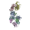
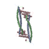
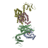
| Title | Structure of the Ca2+-bound Rabphilin 3A C2B domain SNAP25 complex (P21 space group) | ||||||
 Components Components |
| ||||||
 Keywords Keywords | EXOCYTOSIS / membrane fusion / calcium / C2 domain | ||||||
| Function / homology |  Function and homology information Function and homology informationselenium binding / spontaneous neurotransmitter secretion / BLOC-1 complex / calcium-dependent activation of synaptic vesicle fusion / positive regulation of glutamate secretion, neurotransmission / synaptic vesicle fusion to presynaptic active zone membrane / Other interleukin signaling / synaptobrevin 2-SNAP-25-syntaxin-1a-complexin II complex / synaptobrevin 2-SNAP-25-syntaxin-1a complex / synaptobrevin 2-SNAP-25-syntaxin-1a-complexin I complex ...selenium binding / spontaneous neurotransmitter secretion / BLOC-1 complex / calcium-dependent activation of synaptic vesicle fusion / positive regulation of glutamate secretion, neurotransmission / synaptic vesicle fusion to presynaptic active zone membrane / Other interleukin signaling / synaptobrevin 2-SNAP-25-syntaxin-1a-complexin II complex / synaptobrevin 2-SNAP-25-syntaxin-1a complex / synaptobrevin 2-SNAP-25-syntaxin-1a-complexin I complex / presynaptic dense core vesicle exocytosis / extrinsic component of presynaptic membrane / calcium ion-regulated exocytosis of neurotransmitter / Glutamate Neurotransmitter Release Cycle / Norepinephrine Neurotransmitter Release Cycle / Acetylcholine Neurotransmitter Release Cycle / Serotonin Neurotransmitter Release Cycle / GABA synthesis, release, reuptake and degradation / Dopamine Neurotransmitter Release Cycle / cholinergic synapse / short-term synaptic potentiation / extrinsic component of synaptic vesicle membrane / positive regulation of calcium ion-dependent exocytosis / regulation of establishment of protein localization / ribbon synapse / positive regulation of vesicle fusion / SNARE complex / SNAP receptor activity / calcium-dependent phospholipid binding / positive regulation of synaptic plasticity / inositol 1,4,5 trisphosphate binding / positive regulation of hormone secretion / neurotransmitter secretion / dendritic spine organization / regulation of NMDA receptor activity / regulation of synaptic vesicle cycle / extrinsic component of membrane / syntaxin-1 binding / insulin secretion / Neutrophil degranulation / endosomal transport / myosin binding / regulation of synapse assembly / regulation of neuron projection development / phosphate ion binding / SNARE complex assembly / synaptic vesicle priming / exocytosis / synaptic vesicle exocytosis / associative learning / voltage-gated potassium channel activity / axonal growth cone / long-term memory / phosphatidylinositol-4,5-bisphosphate binding / photoreceptor inner segment / somatodendritic compartment / voltage-gated potassium channel complex / axonogenesis / establishment of localization in cell / secretory granule / SNARE binding / filopodium / neuromuscular junction / trans-Golgi network / locomotory behavior / intracellular protein transport / phospholipid binding / small GTPase binding / neuron differentiation / positive regulation of insulin secretion / calcium-dependent protein binding / terminal bouton / synaptic vesicle / long-term synaptic potentiation / lamellipodium / actin cytoskeleton / synaptic vesicle membrane / growth cone / presynaptic membrane / cell cortex / vesicle / dendritic spine / cytoskeleton / transmembrane transporter binding / postsynaptic membrane / endosome / neuron projection / protein domain specific binding / axon / neuronal cell body / calcium ion binding / lipid binding / synapse / protein-containing complex binding / perinuclear region of cytoplasm / glutamatergic synapse / protein-containing complex / zinc ion binding / membrane / plasma membrane Similarity search - Function | ||||||
| Biological species |  | ||||||
| Method |  X-RAY DIFFRACTION / SYNCHROTRON / MOLECULAR REPLACEMENT / Resolution: 2.8 Å X-RAY DIFFRACTION / SYNCHROTRON / MOLECULAR REPLACEMENT / Resolution: 2.8 Å | ||||||
 Authors Authors | Verdaguer, N. / Ferrer-Orta, C. | ||||||
 Citation Citation | Journal: Proc. Natl. Acad. Sci. U.S.A. / Year: 2017 Title: Structural characterization of the Rabphilin-3A-SNAP25 interaction. Authors: Ferrer-Orta, C. / Perez-Sanchez, M.D. / Coronado-Parra, T. / Silva, C. / Lopez-Martinez, D. / Baltanas-Copado, J. / Gomez-Fernandez, J.C. / Corbalan-Garcia, S. / Verdaguer, N. | ||||||
| History |
|
- Structure visualization
| Structure viewer | Molecule: MolmilJmol/JSmol |
|---|
- Downloads & links
-Download
| PDBx/mmCIF format | 5low.cif.gz | 289.1 KB | Display | PDBx/mmCIF format |
|---|---|---|---|---|
| PDB format | pdb5low.ent.gz | 231.2 KB | Display | PDB format |
| PDBx/mmJSON format | 5low.json.gz | Tree view | PDBx/mmJSON format | |
| Others |  Other downloads Other downloads |
-Validation report
| Arichive directory | https://data.pdbj.org/pub/pdb/validation_reports/lo/5lowftp://data.pdbj.org/pub/pdb/validation_reports/lo/5low | HTTPS FTP |
|---|
-Related structure data
| Related structure data |  5lo8C  5lobSC S: Starting model for refinement C: citing same article ( |
|---|---|
| Similar structure data |


-Links
 PDBj
PDBj


- Assembly
| Deposited unit | 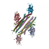
| ||||||||
|---|---|---|---|---|---|---|---|---|---|
| 1 | 
| ||||||||
| 2 | 
| ||||||||
| Unit cell |
|
-Components
-Protein , 1 types, 6 molecules ABCHIJ
| #1: Protein | Mass: 18579.307 Da / Num. of mol.: 6 Source method: isolated from a genetically manipulated source Source: (gene. exp.)  |
|---|
-Synaptosomal-associated protein ... , 2 types, 8 molecules DFKMEGLN
| #2: Protein | Mass: 10841.079 Da / Num. of mol.: 4 Source method: isolated from a genetically manipulated source Source: (gene. exp.) #3: Protein | Mass: 9223.266 Da / Num. of mol.: 4 Source method: isolated from a genetically manipulated source Source: (gene. exp.) |
|---|
-Non-polymers , 3 types, 113 molecules 


| #4: Chemical | ChemComp-GOL /  Mass: 92.094 Da / Num. of mol.: 7 / Source method: obtained synthetically / Formula: C3H8O3 Mass: 92.094 Da / Num. of mol.: 7 / Source method: obtained synthetically / Formula: C3H8O3#5: Chemical | ChemComp-CA /  Mass: 40.078 Da / Num. of mol.: 12 / Source method: obtained synthetically / Formula: Ca Mass: 40.078 Da / Num. of mol.: 12 / Source method: obtained synthetically / Formula: Ca#6: Water | ChemComp-HOH / | Mass: 18.015 Da / Num. of mol.: 94 / Source method: isolated from a natural source / Formula: H2O |
|---|
-Experimental details
-Experiment
| Experiment | Method: X-RAY DIFFRACTION / Number of used crystals: 1 |
|---|
- Sample preparation
| Crystal | Density Matthews: 2.43 Å3/Da / Density % sol: 49.48 % |
|---|---|
| Crystal grow | Temperature: 293 K / Method: vapor diffusion, hanging drop / pH: 8.5 Details: 20% PEG3350 0.35 M Ammonium sulfate 0.1 M Tris (pH 8.5) al's oil |
-Data collection
| Diffraction | Mean temperature: 100 K | |||||||||||||||
|---|---|---|---|---|---|---|---|---|---|---|---|---|---|---|---|---|
| Diffraction source | Source: SYNCHROTRON / Site: ESRF  / Beamline: ID23-2 / Wavelength: 0.8726 Å / Beamline: ID23-2 / Wavelength: 0.8726 Å | |||||||||||||||
| Detector | Type: DECTRIS PILATUS 6M / Detector: PIXEL / Date: May 14, 2015 | |||||||||||||||
| Radiation | Protocol: SINGLE WAVELENGTH / Monochromatic (M) / Laue (L): M / Scattering type: x-ray | |||||||||||||||
| Radiation wavelength | Wavelength: 0.8726 Å / Relative weight: 1 | |||||||||||||||
| Reflection twin |
| |||||||||||||||
| Reflection | Resolution: 2.8→47.9 Å / Num. obs: 43944 / % possible obs: 98.3 % / Redundancy: 2.4 % / Rmerge(I) obs: 0.173 / Net I/σ(I): 4.8 | |||||||||||||||
| Reflection shell | Resolution: 2.8→2.95 Å / Redundancy: 2.4 % / % possible all: 96.2 |
- Processing
| Software |
| ||||||||||||||||||||||||||||||||||||||||||||||||||||||||||||||||||||||||||||||||||||||||||||||||||||||||||||||||||||||||||||||||||||||||||||||||||||||||||||||||||||||||||||||||||||||
|---|---|---|---|---|---|---|---|---|---|---|---|---|---|---|---|---|---|---|---|---|---|---|---|---|---|---|---|---|---|---|---|---|---|---|---|---|---|---|---|---|---|---|---|---|---|---|---|---|---|---|---|---|---|---|---|---|---|---|---|---|---|---|---|---|---|---|---|---|---|---|---|---|---|---|---|---|---|---|---|---|---|---|---|---|---|---|---|---|---|---|---|---|---|---|---|---|---|---|---|---|---|---|---|---|---|---|---|---|---|---|---|---|---|---|---|---|---|---|---|---|---|---|---|---|---|---|---|---|---|---|---|---|---|---|---|---|---|---|---|---|---|---|---|---|---|---|---|---|---|---|---|---|---|---|---|---|---|---|---|---|---|---|---|---|---|---|---|---|---|---|---|---|---|---|---|---|---|---|---|---|---|---|---|
| Refinement | Method to determine structure: MOLECULAR REPLACEMENT Starting model: 5LOB Resolution: 2.8→45.03 Å / Cor.coef. Fo:Fc: 0.873 / Cor.coef. Fo:Fc free: 0.851 / SU B: 8.482 / SU ML: 0.183 / Cross valid method: THROUGHOUT / ESU R Free: 0.085 / Stereochemistry target values: MAXIMUM LIKELIHOOD / Details: HYDROGENS HAVE BEEN ADDED IN THE RIDING POSITIONS
| ||||||||||||||||||||||||||||||||||||||||||||||||||||||||||||||||||||||||||||||||||||||||||||||||||||||||||||||||||||||||||||||||||||||||||||||||||||||||||||||||||||||||||||||||||||||
| Solvent computation | Ion probe radii: 0.8 Å / Shrinkage radii: 0.8 Å / VDW probe radii: 1.2 Å / Solvent model: MASK | ||||||||||||||||||||||||||||||||||||||||||||||||||||||||||||||||||||||||||||||||||||||||||||||||||||||||||||||||||||||||||||||||||||||||||||||||||||||||||||||||||||||||||||||||||||||
| Displacement parameters | Biso mean: 46.33 Å2
| ||||||||||||||||||||||||||||||||||||||||||||||||||||||||||||||||||||||||||||||||||||||||||||||||||||||||||||||||||||||||||||||||||||||||||||||||||||||||||||||||||||||||||||||||||||||
| Refinement step | Cycle: LAST / Resolution: 2.8→45.03 Å
| ||||||||||||||||||||||||||||||||||||||||||||||||||||||||||||||||||||||||||||||||||||||||||||||||||||||||||||||||||||||||||||||||||||||||||||||||||||||||||||||||||||||||||||||||||||||
| Refine LS restraints |
|