 Movie
Movie Controller


+ Open data

- Basic information
| Entry | Database: PDB / ID: 3tj0 | ||||||
|---|---|---|---|---|---|---|---|
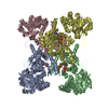
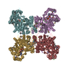
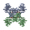
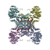
| Title | Crystal Structure of Influenza B Virus Nucleoprotein | ||||||
 Components Components | Nucleoprotein | ||||||
 Keywords Keywords | RNA BINDING PROTEIN / RNA-binding / Homo-oligomerization / Transcription regulation / Nucleus | ||||||
| Function / homology |  Function and homology information Function and homology informationhelical viral capsid / viral penetration into host nucleus / host cell / viral nucleocapsid / ribonucleoprotein complex / symbiont entry into host cell / host cell nucleus / structural molecule activity / RNA binding Similarity search - Function | ||||||
| Biological species |  Influenza B virus Influenza B virus | ||||||
| Method |  X-RAY DIFFRACTION / SYNCHROTRON / MOLECULAR REPLACEMENT / Resolution: 3.233 Å X-RAY DIFFRACTION / SYNCHROTRON / MOLECULAR REPLACEMENT / Resolution: 3.233 Å | ||||||
 Authors Authors | Ng, A.K.L. / Zhang, H. / Liu, J. / Au, S.W.N. / Wang, J. / Shaw, P.C. | ||||||
 Citation Citation | Journal: J.Virol. / Year: 2012 Title: Structural basis for RNA binding and homo-oligomer formation by influenza B virus nucleoprotein Authors: Ng, A.K.L. / Lam, M.K.H. / Zhang, H. / Liu, J. / Au, S.W.N. / Chan, P.K.S. / Wang, J. / Shaw, P.C. | ||||||
| History |
|
- Structure visualization
| Structure viewer | Molecule: MolmilJmol/JSmol |
|---|
- Downloads & links
-Download
| PDBx/mmCIF format | 3tj0.cif.gz | 183.8 KB | Display | PDBx/mmCIF format |
|---|---|---|---|---|
| PDB format | pdb3tj0.ent.gz | 145.7 KB | Display | PDB format |
| PDBx/mmJSON format | 3tj0.json.gz | Tree view | PDBx/mmJSON format | |
| Others |  Other downloads Other downloads |
-Validation report
| Arichive directory | https://data.pdbj.org/pub/pdb/validation_reports/tj/3tj0ftp://data.pdbj.org/pub/pdb/validation_reports/tj/3tj0 | HTTPS FTP |
|---|
-Related structure data
| Related structure data | 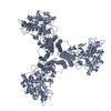 2q06S S: Starting model for refinement |
|---|---|
| Similar structure data |

-Links
 PDBj
PDBj- Assembly
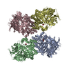
| Deposited unit | 
| ||||||||
|---|---|---|---|---|---|---|---|---|---|
| 1 | 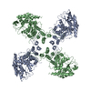
| ||||||||
| 2 | 
| ||||||||
| Unit cell |
|
-Components
| #1: Protein | Mass: 61882.129 Da / Num. of mol.: 2 Source method: isolated from a genetically manipulated source Source: (gene. exp.) Influenza B virus / Strain: B/Managua/4577.01/2008 / Gene: NP / Production host:  Sequence details | THIS SEQUENCE IS THE NATURAL POLYMORPHI | |
|---|
-Experimental details
-Experiment
| Experiment | Method: X-RAY DIFFRACTION / Number of used crystals: 1 |
|---|
- Sample preparation
| Crystal | Density Matthews: 2.64 Å3/Da / Density % sol: 53.39 % |
|---|---|
| Crystal grow | Temperature: 289 K / Method: vapor diffusion, hanging drop / pH: 7 Details: 1.4M sodium-potassium phosphate, pH 7, VAPOR DIFFUSION, HANGING DROP, temperature 289K |
-Data collection
| Diffraction | Mean temperature: 100 K |
|---|---|
| Diffraction source | Source: SYNCHROTRON / Site: SSRF  / Beamline: BL17U / Wavelength: 0.97941 Å / Beamline: BL17U / Wavelength: 0.97941 Å |
| Detector | Type: ADSC QUANTUM 315r / Detector: CCD / Date: May 18, 2011 |
| Radiation | Protocol: SINGLE WAVELENGTH / Monochromatic (M) / Laue (L): M / Scattering type: x-ray |
| Radiation wavelength | Wavelength: 0.97941 Å / Relative weight: 1 |
| Reflection | Resolution: 3.2→50 Å / Num. all: 21367 / Num. obs: 19883 / % possible obs: 93.1 % / Observed criterion σ(F): 0 / Observed criterion σ(I): 0 |
| Reflection shell | Resolution: 3.23→3.35 Å / Redundancy: 7.3 % / Rmerge(I) obs: 0.387 / Mean I/σ(I) obs: 2.9 / Num. unique all: 1998 / % possible all: 95.1 |
- Processing
| Software |
| ||||||||||||||||||||||||||||||||||||||||||||||||
|---|---|---|---|---|---|---|---|---|---|---|---|---|---|---|---|---|---|---|---|---|---|---|---|---|---|---|---|---|---|---|---|---|---|---|---|---|---|---|---|---|---|---|---|---|---|---|---|---|---|
| Refinement | Method to determine structure: MOLECULAR REPLACEMENT Starting model: 2Q06 Resolution: 3.233→47.055 Å / SU ML: 0.41 / σ(F): 0 / Phase error: 31.88 / Stereochemistry target values: ML
| ||||||||||||||||||||||||||||||||||||||||||||||||
| Solvent computation | Shrinkage radii: 0.16 Å / VDW probe radii: 0.5 Å / Solvent model: FLAT BULK SOLVENT MODEL / Bsol: 47.073 Å2 / ksol: 0.343 e/Å3 | ||||||||||||||||||||||||||||||||||||||||||||||||
| Displacement parameters |
| ||||||||||||||||||||||||||||||||||||||||||||||||
| Refinement step | Cycle: LAST / Resolution: 3.233→47.055 Å
| ||||||||||||||||||||||||||||||||||||||||||||||||
| Refine LS restraints |
| ||||||||||||||||||||||||||||||||||||||||||||||||
| LS refinement shell | Refine-ID: X-RAY DIFFRACTION / Total num. of bins used: 7
|
